 Indietro
Hemostasis and Blood: Structure, Function, and Disorders
Guida di studio - Note intelligenti
 Appunti personalizzati basati sui tuoi materiali, ampliati con definizioni chiave, esempi e contesto.
Hemostasis and Blood Composition
Overview of Blood
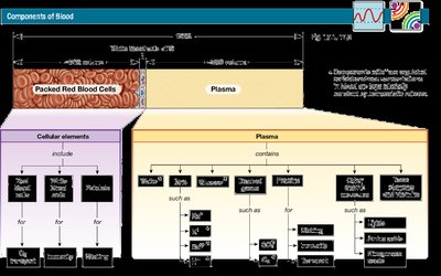
Blood is a specialized connective tissue that plays a vital role in transport, regulation, and protection within the human body. It consists of a liquid matrix called plasma and various formed elements, including red blood cells, white blood cells, and platelets.
Plasma: The fluid matrix of blood, making up about 55% of total blood volume. It is primarily water but also contains proteins, ions, nutrients, gases, and waste products.
Formed Elements: These include erythrocytes (red blood cells), leukocytes (white blood cells), and platelets (cell fragments).
Plasma Components
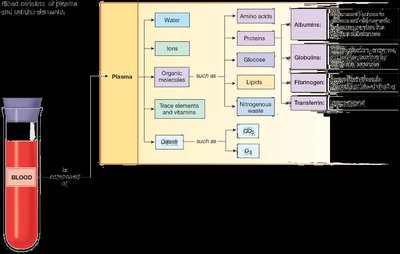
Plasma is the extracellular matrix of blood and is similar in composition to interstitial fluid, but it contains unique plasma proteins.
Water: ~92% of plasma; serves as a solvent and medium for transport.
Proteins: ~7% of plasma; includes albumins (osmotic balance), globulins (immune function), fibrinogen (clotting), and transferrin (iron transport).
Other Solutes: Ions (Na+, K+, Cl-, Ca2+, HCO3-), organic molecules (glucose, amino acids, lipids), gases (O2, CO2), trace elements, and vitamins.
Formed Elements of Blood
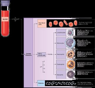
The cellular elements of blood are responsible for oxygen transport, immune defense, and clotting.
Red Blood Cells (Erythrocytes): Transport oxygen and carbon dioxide.
White Blood Cells (Leukocytes): Defend against pathogens. Subtypes include lymphocytes, monocytes, neutrophils, eosinophils, and basophils.
Platelets (Thrombocytes): Cell fragments essential for blood clotting.
Hematopoiesis: Blood Cell Formation
Sites and Regulation of Hematopoiesis
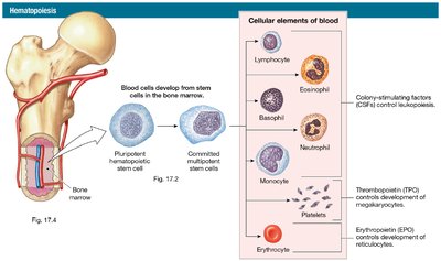
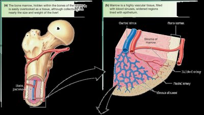
Hematopoiesis is the process of blood cell production, occurring primarily in the red bone marrow. It is regulated by various cytokines and growth factors.
Red Bone Marrow: Active site of blood cell production; contains hematopoietic stem cells.
Yellow Bone Marrow: Contains adipose tissue; inactive in hematopoiesis.
Regulatory Factors: Colony-stimulating factors (CSFs) regulate leukocyte production, thrombopoietin (TPO) regulates platelet production, and erythropoietin (EPO) regulates erythrocyte production.
Bone Marrow Structure
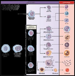
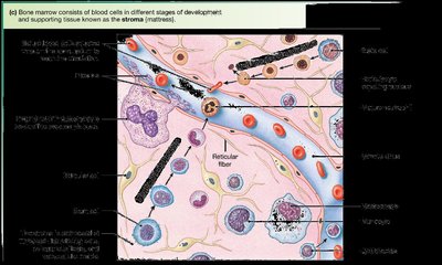
Bone marrow is a highly vascular tissue found within bones, supporting the development of blood cells at various stages.
Stroma: The supportive tissue of bone marrow, consisting of fibroblasts, adipocytes, and blood vessels.
Developmental Stages: Blood cells mature in the marrow before entering circulation.
Red Blood Cells (Erythrocytes)
Structure and Function
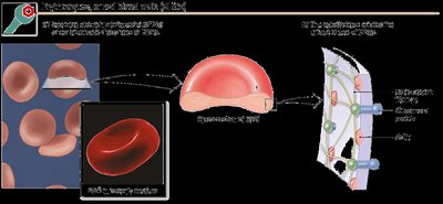
Red blood cells are biconcave, anucleate cells specialized for gas transport. Their shape increases surface area for efficient gas exchange and allows flexibility in capillaries.
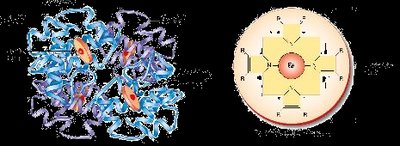
Hemoglobin: The oxygen-carrying protein in RBCs, composed of four globin chains and four heme groups, each containing an iron atom.
Lifespan: Approximately 120 days; old RBCs are removed by the spleen and liver.
Hemoglobin Synthesis and Iron Metabolism
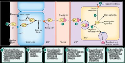
Hemoglobin synthesis requires iron, which is absorbed from the diet and transported by transferrin. Excess iron is stored in the liver as ferritin.
Iron Cycle: Iron is recycled from old RBCs and reused for new hemoglobin synthesis.
Bilirubin: The breakdown product of heme, excreted in bile and urine. Elevated bilirubin causes jaundice.
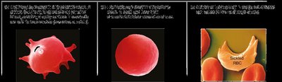
Red Blood Cell Disorders
Disorders affecting RBCs can impair oxygen transport and cause clinical symptoms.
Anemia: Reduced oxygen-carrying capacity due to low hemoglobin or RBC count. Causes include blood loss, hemolysis, and decreased production.
Polycythemia: Excess RBCs, increasing blood viscosity.
Hemolytic Anemia: Premature destruction of RBCs (e.g., sickle cell disease, hereditary spherocytosis).
Platelets and Hemostasis
Platelet Structure and Function
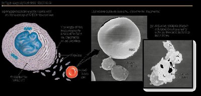
Platelets are small, anucleate cell fragments derived from megakaryocytes. They play a crucial role in hemostasis, immunity, and inflammation.
Lifespan: About 10 days.
Granules: Contain cytokines and growth factors essential for clotting and tissue repair.
Hemostasis: Prevention of Blood Loss
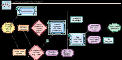
Hemostasis is the physiological process that stops bleeding at the site of injury while maintaining blood flow elsewhere in the circulation. It involves three major steps:
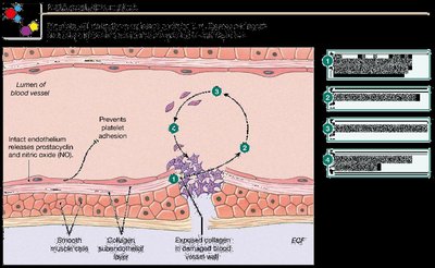
Vasoconstriction: Immediate constriction of damaged blood vessels to reduce blood flow.
Platelet Plug Formation: Platelets adhere to exposed collagen, become activated, and aggregate to form a temporary plug.
Coagulation Cascade: A series of enzymatic reactions leading to the conversion of fibrinogen to fibrin, stabilizing the platelet plug into a clot.
Coagulation Pathways
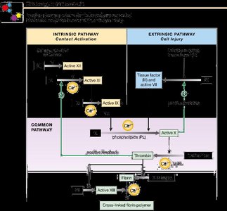
Coagulation involves two converging pathways:
Intrinsic Pathway (Contact Activation): Initiated by damage inside the vessel.
Extrinsic Pathway (Cell Injury): Triggered by external trauma exposing tissue factor.
Common Pathway: Both pathways lead to the activation of thrombin, which converts fibrinogen to fibrin.
Fibrinolysis and Anticoagulation
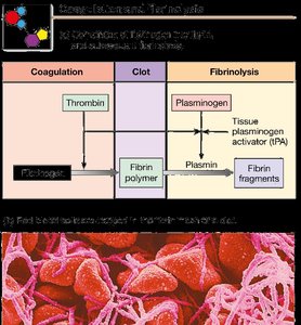
After vessel repair, the clot is dissolved by fibrinolysis. Anticoagulants prevent excessive clotting and maintain blood fluidity.
Fibrinolysis: Plasminogen is activated to plasmin, which digests fibrin and dissolves the clot.
Endogenous Anticoagulants: Heparin, antithrombin III, and protein C inhibit various steps in the coagulation cascade.
Hemophilia: A group of disorders characterized by defective or missing coagulation factors, leading to ineffective clotting.
Complete Blood Count (CBC) and Normal Ranges
Key Laboratory Values
A CBC provides quantitative information about the formed elements of blood and is essential for diagnosing various hematological conditions.
Test | Males | Females | Description |
|---|---|---|---|
Hematocrit (%) | 40–54 | 37–47 | Percentage of blood volume occupied by RBCs |
Hemoglobin (g/dL) | 14–17 | 12–16 | Oxygen-carrying capacity |
RBC Count (cells/mL) | 4.5–6.5 × 10⁶ | 3.9–5.6 × 10⁶ | Number of erythrocytes |
Total WBC Count (cells/mL) | 4–11 × 104 | All leukocytes | |
Platelets (per µL) | 150–450 × 104 | Clotting ability | |
Differential WBC Count:
Leukocyte Type | Normal Range (%) |
|---|---|
Neutrophils | 50–70 |
Eosinophils | 1–4 |
Basophils | <1 |
Lymphocytes | 20–40 |
Monocytes | 2–8 |
Summary Table: Key Factors in Coagulation and Fibrinolysis
Factor | Source | Role |
|---|---|---|
Collagen | Subendothelial ECM | Starts intrinsic pathway |
von Willebrand factor | Endothelium, megakaryocytes | Regulates factor VIII, platelet adhesion |
Tissue factor (III) | Most cells except platelets | Starts extrinsic pathway |
Prothrombin/thrombin (II) | Liver, plasma | Converts fibrinogen to fibrin |
Fibrinogen/fibrin (I) | Liver, plasma | Forms insoluble fibers |
Plasminogen/plasmin | Liver, plasma | Dissolves fibrin (fibrinolysis) |
Antithrombin III | Liver, plasma | Anticoagulant |
Key Equations
Hematocrit Calculation:
Oxygen Carrying Capacity:
Additional info:
Some images and tables have been logically grouped and expanded for clarity and completeness.
Key terms and processes have been defined and explained for self-contained study.
