 Indietro
Histology and Tissue Types: Structured Study Notes for Anatomy & Physiology
Guida di studio - Note intelligenti
 Appunti personalizzati basati sui tuoi materiali, ampliati con definizioni chiave, esempi e contesto.
Histology: The Study of Tissues
Definition and Major Types of Tissues
Histology is the study of tissues, which are groups of structurally and functionally related cells and their external environment. Tissues perform common functions and are essential for organ structure and function.
Tissue: A group of cells and their ECM performing a specific function.
All tissues have a discrete population of cells and a surrounding extracellular matrix (ECM).
Types of Tissues
Epithelial Tissue: Sheets of tightly packed cells with little ECM; lines body surfaces and cavities.
Connective Tissue: Connects other tissues; ECM is prominent, cells are scattered; functions include binding, support, protection, and transport.
Muscle Tissue: Composed of cells that contract and generate force; little ECM.
Nervous Tissue: Composed of cells that generate, send, and receive messages; includes supportive glial cells.
Organs are made up of two or more tissue types working together.
The Extracellular Matrix (ECM)
Structure and Function
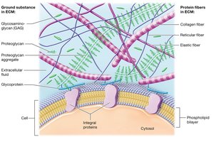
The ECM is composed of substances surrounding the cells in a tissue. It provides structural support, regulates cell behavior, and holds cells in place.
Ground Substance: Gel-like, contains water, ions, nutrients, and other solutes.
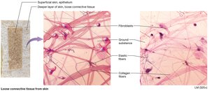
Protein Fibers: Collagen (resists tension), Elastic (stretch and recoil), Reticular (meshwork support).
ECM can be fluid, gel, or solid depending on its components.
Functions: Strength, cell placement, regulation of development, mitotic activity, and survival.
Cell Junctions
Types and Functions
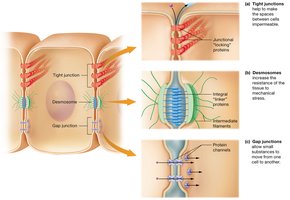
Cell junctions are structures that link neighboring cells, providing mechanical strength and communication.
Tight Junctions: Hold cells tightly, making the space impermeable to macromolecules.
Desmosomes: Hold cells together, allow passage of molecules, distribute mechanical stress.
Gap Junctions: Protein channels allow small substances to pass freely between cells.
Epithelial Tissue
Functions of Epithelial Tissue
Epithelial tissues cover internal and external surfaces, acting as barriers and performing specialized functions.
Protection: Shields underlying tissues.
Immune Defense: Barrier against microorganisms.
Secretion: Forms glands that secrete substances.
Transport: Selectively permeable for nutrient and substance movement.
Sensation: Contains nerves for environmental sensing.
Structure of Epithelial Tissue
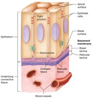
Epithelial tissue is characterized by tightly packed cells, a basement membrane, and an apical surface exposed to the environment or internal cavity.
Classification of Epithelial Cells
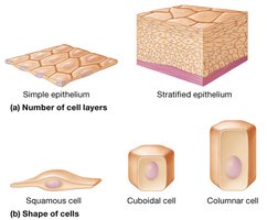
Epithelial cells are classified by the number of cell layers and the shape of cells.
Simple Epithelium: Single layer of cells.
Stratified Epithelium: Multiple layers of cells.
Cell Shapes: Squamous (flat), Cuboidal (cube-shaped), Columnar (tall).
Types of Epithelia
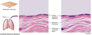
Simple Squamous Epithelium: Thin, flat cells; found in lungs and blood vessels.
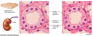
Simple Cuboidal Epithelium: Cube-shaped cells; found in kidney tubules.
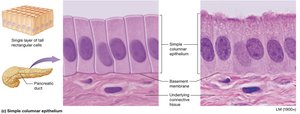
Simple Columnar Epithelium: Tall, rectangular cells; found in digestive tract.
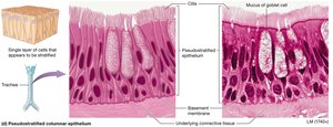
Pseudostratified Columnar Epithelium: Appears stratified but is a single layer; found in respiratory tract.
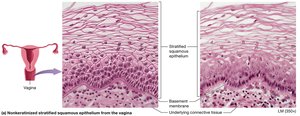
Stratified Squamous Epithelium: Multiple layers; protection; skin, mouth, vagina.
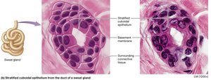
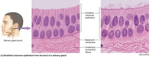
Stratified Cuboidal/Columnar Epithelium: Rare; found in ducts of glands.
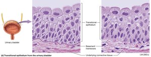
Transitional Epithelium: Specialized for stretching; found in urinary bladder.
Glandular Epithelium
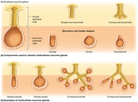
Glands are structures that make and secrete products. They arise from epithelial tissue and are classified as exocrine or endocrine.
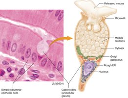
Exocrine Glands: Secrete products via ducts to local surfaces; can be unicellular (e.g., goblet cells) or multicellular.
Endocrine Glands: Secrete hormones directly into the blood; lack ducts.
Exocrine Secretion Types
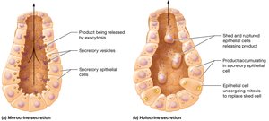
Merocrine Secretion: Products released by exocytosis (e.g., sweat glands).
Holocrine Secretion: Products released when cells rupture and die (e.g., sebaceous glands).
Connective Tissue
Functions and Types
Connective tissue binds, supports, protects, and transports substances. It is divided into connective tissue proper and specialized connective tissue.
Connecting and Binding: Anchors organs and tissues.
Support: Provides structural support (bone, cartilage).
Protection: Shock absorption and organ protection.
Transport: Blood transports substances throughout the body.
Cells of Connective Tissue Proper
Fibroblasts: Produce protein fibers of the ECM.
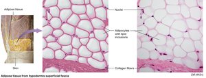
Adipocytes: Fat cells storing lipids.
Mast Cells: Immune cells with inflammatory mediators.
Phagocytes: Immune cells that ingest foreign substances.
Types of Connective Tissue Proper
Loose Connective Tissue: Supports and contains blood vessels and immune cells.
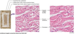
Dense Irregular Connective Tissue: Collagen fibers arranged haphazardly; resists tension in all planes.
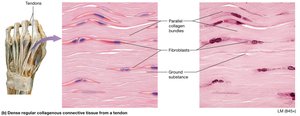
Dense Regular Collagenous Connective Tissue: Parallel collagen fibers; resists tension in one plane.
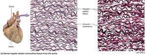
Dense Regular Elastic Connective Tissue: Parallel elastic fibers; found in blood vessels and ligaments.
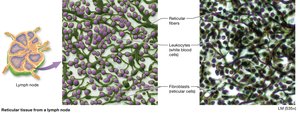
Reticular Tissue: Meshwork of reticular fibers; supports small structures and houses white blood cells.
Adipose Tissue: Fat-storing cells; insulation, protection, energy reserve.
Types of Specialized Connective Tissue
Cartilage: Tough, flexible, absorbs shock; found in joints, ears, nose, respiratory passages.
Bone (Osseous) Tissue: Provides structural support and protection.
Blood: Fluid ECM (plasma); transports oxygen, immune cells, and platelets.
Classes of Cartilage
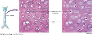
Hyaline Cartilage: Covers ends of bones at joints; most abundant.
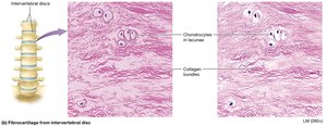
Fibrocartilage: Found in intervertebral discs; strong and elastic.
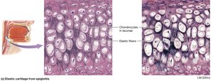
Elastic Cartilage: Found in external ear and larynx; flexible and vibration-sensitive.
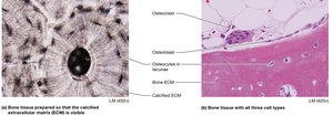
The Cells of Bone (Osseous) Tissue
Osteoblasts: Build bone via deposition.
Osteocytes: Mature bone cells in lacunae; maintain bone.
Osteoclasts: Break down bone via resorption.
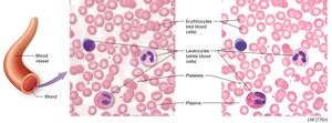
Blood
Erythrocytes (Red Blood Cells): Transport oxygen.
Leukocytes (White Blood Cells): Immune function.
Platelets: Blood clotting.
Muscle Tissue
Structure and Function
Muscle tissue is specialized for contraction and movement. Muscle cells (myocytes) are excitable and contain myofilaments for contraction.
Striated Muscle Cells: Alternating light and dark bands (striations).
Smooth Muscle Cells: Irregular bundles, no visible striations.
Endomysium: ECM surrounding each muscle cell.
Types of Muscle Tissue
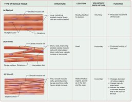
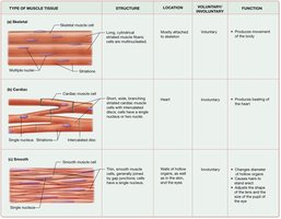
Skeletal Muscle Tissue: Long, striated, multinucleated; voluntary movement.
Cardiac Muscle Tissue: Short, branched, uninucleated; involuntary; intercalated discs for coordinated contraction.
Smooth Muscle Tissue: Flattened, involuntary; found in hollow organs, blood vessels, eyes, skin.
Type | Structure | Location | Voluntary/Involuntary | Function |
|---|---|---|---|---|
Skeletal | Long, striated, multinucleated | Attached to skeleton | Voluntary | Movement |
Cardiac | Short, branched, uninucleated | Heart | Involuntary | Beating of heart |
Smooth | Flattened, no striations | Walls of hollow organs, vessels, eyes, skin | Involuntary | Changes diameter, movement of substances |
Nervous Tissue
Structure and Function
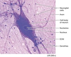
Nervous tissue is the main component of the brain, spinal cord, and nerves. It consists of neurons and neuroglial cells.
Neurons: Excitable cells that send and receive messages; do not undergo mitosis.
Neuroglial Cells: Supportive functions for neurons.
Cell Body (Soma): Contains nucleus and organelles.
Axon: Transmits signals to other cells.
Dendrites: Receive signals from other neurons.
Transport Across Membranes
Passive and Active Transport
Cells transport substances across membranes via passive and active mechanisms. Passive transport does not require energy, while active transport does.
Simple Diffusion: Movement of molecules from high to low concentration.
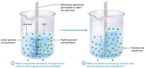
Osmosis: Movement of water across a selectively permeable membrane.
Facilitated Diffusion: Movement of molecules via protein channels.
Pinocytosis: Active transport; cell engulfs fluid.
Osmosis is illustrated as water moving across a membrane into a solution with higher solute concentration, continuing until equilibrium is reached.
Summary Table: Muscle Tissue Types
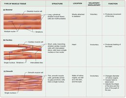
Type of Muscle Tissue | Structure | Location | Voluntary/Involuntary | Function |
|---|---|---|---|---|
Skeletal | Long, striated, multinucleated | Mostly attached to skeleton | Voluntary | Produces movement of the body |
Cardiac | Short, thick, branched, uninucleated | Heart | Involuntary | Produces beating of the heart |
Smooth | Flattened, single nucleus, no striations | Walls of hollow organs, blood vessels, eyes, skin | Involuntary | Changes diameter of tubes, moves substances, adjusts pupil size |
Additional info: Academic context was added to clarify tissue functions, cell types, and structural hierarchy. All images included are directly relevant to the adjacent explanations and reinforce key concepts in histology and tissue structure.
