 Indietro
Introduction to Histology: The Four Primary Tissue Types
Guida di studio - Note intelligenti
 Appunti personalizzati basati sui tuoi materiali, ampliati con definizioni chiave, esempi e contesto.
Introduction to Tissues
Definition and Components
Tissues are groups of structurally and functionally related cells and their external environment that together perform common functions. All tissues share two basic components:
Cells: Related in structure and function.
Extracellular Matrix (ECM): The composition of the ECM differs in each tissue type and provides structural and biochemical support to the surrounding cells.
There are four primary tissue types in the human body:
Epithelial Tissues
Connective Tissues
Muscular Tissues
Nervous Tissues
Epithelial Tissue
Structure and Classification
Epithelial tissue consists of densely packed cells built on top of a basement membrane. It is classified based on the number of cell layers and the shape of the cells:
Simple Epithelium: A single layer of cells attached to the basement membrane.
Stratified Epithelium: Two or more layers of cells; only the basal layer is attached to the basement membrane.
Cell Shapes:
Squamous: Flat cells
Cuboidal: Cube-shaped or spherical cells
Columnar: Tall and skinny cells
Locations and Functions
Epithelial tissues are found:
Covering body surfaces (e.g., skin)
Lining passageways (e.g., blood vessels, kidney tubules, digestive organs, alveoli in lungs)
Lining body cavities (e.g., serous membranes)
Forming glands
Functions include:
Diffusion (e.g., alveoli in lungs)
Absorption/Secretion (e.g., kidney tubules, digestive organs, glands)
Protection (e.g., skin, digestive organs)
Types of Epithelial Tissue
Simple Squamous
Simple Cuboidal
Simple Columnar
Pseudostratified
Stratified Squamous
Stratified Cuboidal
Stratified Columnar
Transitional
Example: Simple squamous epithelium is found in the alveoli of the lungs, facilitating gas exchange.
Connective Tissue
Structure and Fiber Types
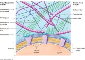
Connective tissue is widely distributed throughout the body and can be comprised of cells, fibers, and ECM. The composition and types of cells and fibers vary between different types of connective tissue. The main fiber types are:
Collagen: Thick fibers that allow tissues to resist tension (pulling and stretching forces).
Reticular: Thin, short fibers that form meshwork or scaffolding to support the cells of connective tissue.
Elastic: Allows tissues to stretch and recoil.
Classes and Functions
Connective Tissue Proper: Loose and dense connective tissue.
Supportive Connective Tissue: Bone and cartilage.
Fluid Connective Tissue: Blood and lymph.
Functions include:
Connecting and binding
Support
Protection
Transportation
Types of Connective Tissue
Loose Connective Tissue: Areolar, Reticular, Adipose
Dense Connective Tissue: Dense Regular, Dense Irregular, Elastic
Fluid Connective Tissue: Blood, Lymph
Supportive Connective Tissue: Compact Bone, Spongy Bone, Cartilage (Hyaline, Elastic, Fibrocartilage)
Example: Dense regular connective tissue forms tendons, which connect muscle to bone.
Muscle Tissue
Structure and Types
Muscle tissue is comprised of muscle cells (myocytes or muscle fibers) that are excitable and respond to electrical or chemical stimulation. The primary function of muscle tissue is to produce movement.
Skeletal Muscle: Attached to bones; voluntary and striated.
Cardiac Muscle: Found only in the heart; involuntary and striated.
Smooth Muscle: Found in the walls of organs; involuntary and non-striated.
Characteristics
Striated/Non-Striated: Striated muscle (skeletal and cardiac) has alternating light-dark bands called striations. Smooth muscle is non-striated.
Voluntary/Involuntary: Skeletal muscle is voluntary; cardiac and smooth muscle are involuntary.
Example: Cardiac muscle contracts rhythmically to pump blood throughout the body.
Nervous Tissue
Structure and Function
Nervous tissue makes up the majority of the brain, spinal cord, and nerves. Its primary function is communication.
Neurons: Contain a cell body with dendrites and an axon; generate, conduct, and receive nerve impulses.
Neuroglial Cells (Glia): Six different types that perform various functions, including supporting neurons, monitoring the composition of the extracellular fluid, and circulating fluid around the brain and spinal cord.
Example: Neurons transmit electrical signals that allow for rapid communication between different parts of the body.
Membranes
Structure and Functions
A membrane is a thin sheet of one or more tissues that lines a body surface or cavity. It consists of a superficial layer of secretory cells and a layer of connective tissue.
Anchors organs in place
Serves as barriers
Functions in immunity
Secretes various substances
Types of Membranes
Serous Membrane
Synovial Membrane
Mucous Membrane
Cutaneous Membrane
Example: The serous membrane lines body cavities and secretes serous fluid to reduce friction between organs.
Additional info: The notes have been expanded to provide academic context, definitions, and examples for each tissue type and membrane. The included image directly illustrates the components of the extracellular matrix in connective tissue, supporting the explanation in the relevant section.
