 Indietro
Introduction to the Nervous System and Nervous Tissue: Structured Study Notes
Guida di studio - Note intelligenti
 Appunti personalizzati basati sui tuoi materiali, ampliati con definizioni chiave, esempi e contesto.
Overview of the Nervous System
Functions of the Nervous System
The nervous system is responsible for controlling perception, voluntary movement, consciousness, personality, learning, and memory. It works closely with the endocrine system to maintain homeostasis, regulating variables such as respiratory rate, blood pressure, body temperature, sleep/wake cycle, and blood pH.
Perception and Experience: Directs how we interpret and respond to our environment.
Voluntary Movement: Initiates and controls muscle activity.
Homeostasis: Maintains internal balance with the endocrine system.
Structural Divisions of the Nervous System
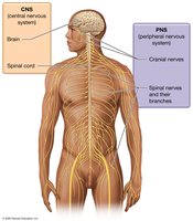
The nervous system is divided into two main structural components: the Central Nervous System (CNS) and the Peripheral Nervous System (PNS).
CNS: Composed of the brain and spinal cord. The brain contains about 100 billion neurons, while the spinal cord contains about 100 million neurons.
PNS: Consists of cranial nerves (12 pairs) and spinal nerves (31 pairs) and their branches. Nerves are bundles of long neuron axons surrounded by connective tissue.
Functional Divisions of the Nervous System
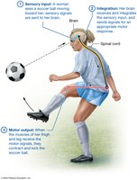
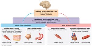
Functionally, the nervous system is divided into sensory, integrative, and motor divisions.
Sensory (Afferent) Division: Gathers information from internal and external environments via sensory receptors.
Integrative Functions: CNS analyzes and interprets sensory input, determining appropriate responses.
Motor (Efferent) Division: Executes actions in response to integration, such as muscle contractions and glandular secretion.
Nervous Tissue
Cellular Composition
Nervous tissue consists of approximately 80% cells and 20% extracellular matrix. The two main cell types are neurons and neuroglial cells.
Neurons: Excitable cells responsible for transmitting electrical signals.
Neuroglial Cells: Supportive cells that maintain the environment, protect neurons, and assist in their functions.
Neurons
Structure and Function
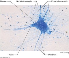
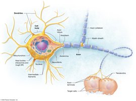
Neurons are specialized for sending and receiving signals in the form of action potentials. They are generally long-lived and amitotic. Most neurons have a central cell body (soma), dendrites, and a single axon.
Cell Body (Soma): Contains most organelles, including abundant ribosomes and rough ER (Nissl bodies), Golgi apparatus, nucleoli, mitochondria, and cytoskeleton (neurofibrils and microtubules).
Dendrites: Short, highly branched processes that receive input and transmit electrical signals toward the cell body.
Axon: Long process that carries electrical signals away from the cell body. Includes axon hillock, axon collaterals, telodendria, and axon terminals (synaptic knobs).
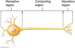
Functional Regions of Neurons
Receptive Region: Dendrites and cell body receive signals.
Conducting Region: Axon transmits signals.
Secretory Region: Axon terminals release neurotransmitters to target cells.
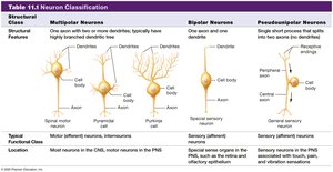
Structural Classification of Neurons
Structural Class | Features | Function |
|---|---|---|
Multipolar | Single axon, multiple dendrites | Motor neurons, interneurons |
Bipolar | One axon, one dendrite | Sensory neurons (retina, olfactory epithelium) |
Pseudounipolar | Single axon with two branches | Sensory neurons (touch, pain) |
Functional Classification of Neurons
Sensory (Afferent) Neurons: Carry signals toward the CNS; detect input from internal and external environments.
Interneurons: Relay messages within the CNS; most abundant.
Motor (Efferent) Neurons: Carry signals away from the CNS to muscles and glands.
Groups of Neuron Cell Bodies and Axons
Nuclei: Clusters of cell bodies in the CNS.
Ganglia: Clusters of cell bodies in the PNS.
Tracts: Bundles of axons in the CNS.
Nerves: Bundles of axons in the PNS.
Neuroglia (Neuroglial Cells)
Types and Functions
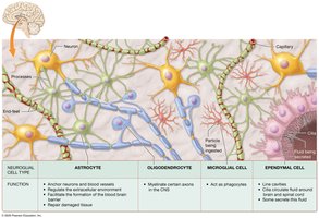
Neuroglia support, protect, and maintain the environment around neurons. They can undergo mitosis and fill gaps when neurons die. There are six types: four in the CNS and two in the PNS.
Astrocytes: Anchor neurons and blood vessels, regulate extracellular environment, assist in blood-brain barrier formation, and repair damaged tissue.
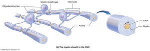
Oligodendrocytes: Form myelin sheath in CNS.
Microglia: Act as phagocytes, ingest debris, stimulate inflammation.
Ependymal Cells: Circulate and monitor cerebrospinal fluid.
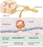
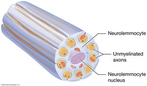
Neurolemmocytes (Schwann Cells): Myelinate axons in PNS, assist in repair.
Satellite Cells: Surround and support cell bodies in PNS.
The Myelin Sheath
Structure and Function
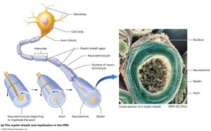
The myelin sheath is formed by neurolemmocytes in the PNS and oligodendrocytes in the CNS. It consists of repeating layers of plasma membrane, acting as an insulator and increasing the speed of nerve impulse conduction.
Myelinated Axons: Conduct action potentials 15–150 times faster than unmyelinated axons.
Internodes: Segments of axon covered by myelin.
Nodes of Ranvier: Gaps between internodes.
Regeneration of Nervous Tissue
Repair in the PNS
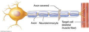
Regeneration is limited in humans, especially in the CNS. In the PNS, repair is possible if the cell body remains intact and conditions are ideal.
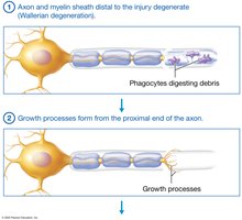
Axon and myelin sheath distal to injury degenerate (Wallerian degeneration).
Phagocytes digest debris.
Growth processes form from the proximal end of the axon.
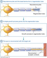
Neurolemmocytes and basal lamina form a regeneration tube.
A single growth process grows into the tube and reconnects with the target cell.
Electrophysiology of Neurons
Resting Membrane Potential
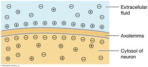
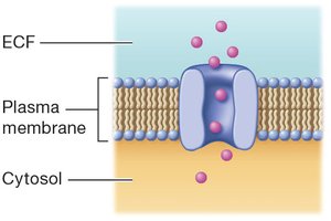
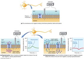
The resting membrane potential is the electrical gradient across the plasma membrane, typically about -70 mV. It is maintained by the movement of potassium ions through leak channels.
Ion Channels and Gradients
Leak Channels: Always open, allow ions to move passively.
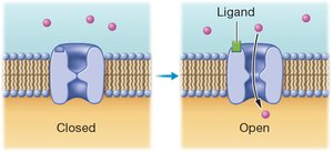
Ligand-Gated Channels: Open in response to chemical binding.
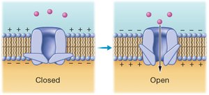
Voltage-Gated Channels: Open/close in response to changes in membrane potential.
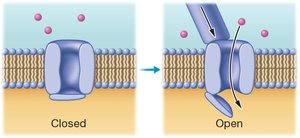
Mechanically Gated Channels: Open/close in response to mechanical stimulation.
Changes in Membrane Potential
Depolarization: Membrane potential becomes less negative (influx of cations).
Repolarization: Return to resting membrane potential.
Hyperpolarization: Membrane potential becomes more negative than resting.
Local and Action Potentials
Local (Graded) Potentials
Small, local changes in membrane potential.
Can cause depolarization or hyperpolarization.
Reversible and decremental (diminish over distance).
Action Potentials
Uniform, rapid depolarization and repolarization.
Generated at the axon’s trigger zone.
All-or-none principle: must reach threshold to occur.
States of Voltage-Gated Channels
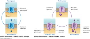
Potassium Channels: Resting (closed) and activated (open).
Sodium Channels: Resting, activated, and inactivated states.
Events of an Action Potential
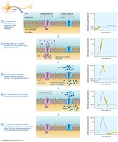
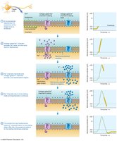
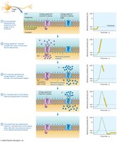
Local potential depolarizes axolemma to threshold (-55 mV).
Voltage-gated sodium channels activate, sodium enters, depolarization to +30 mV.
Sodium channels inactivate, potassium channels activate, repolarization begins.
Sodium channels return to resting state, repolarization continues.
Axolemma may hyperpolarize before returning to resting potential.
The Refractory Period
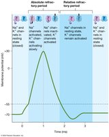
Absolute Refractory Period: No additional action potential can be generated.
Relative Refractory Period: Stronger stimulus required to generate action potential.
Propagation of Action Potentials
Mechanism
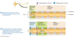
Action potentials are self-propagating, moving in one direction from trigger zone to axon terminals.
Depolarizing current triggers adjacent voltage-gated channels, firing another action potential.
Conduction Speed
Continuous Conduction: Occurs in unmyelinated axons; each section must be depolarized.
Saltatory Conduction: Occurs in myelinated axons; action potential jumps from node to node.
Classification of Axons by Conduction Speed
Type | Diameter | Myelination | Speed | Function |
|---|---|---|---|---|
A | 5–20 µm | Myelinated | ~120 m/s | Sensory/motor to skeletal muscle |
B | 2–3 µm | Myelinated | ~15 m/s | Sensory from organs, ANS efferent |
C | 0.5–1.5 µm | Unmyelinated | ~0.5–2 m/s | Pain, temperature, ANS efferent |
Neuronal Synapses
Types of Synapses
Electrical Synapses: Direct electrical coupling via gap junctions; bidirectional and nearly instantaneous.
Chemical Synapses: Use neurotransmitters; unidirectional, involve synaptic delay, and allow varied effects.
Events at a Chemical Synapse
Action potential triggers calcium channels to open.
Calcium influx causes synaptic vesicles to release neurotransmitters.
Neurotransmitters bind to postsynaptic receptors.
Ion channels open, leading to local potential and possibly action potential.
Postsynaptic Potentials
Excitatory Postsynaptic Potential (EPSP): Local depolarization, moves membrane potential closer to threshold.
Inhibitory Postsynaptic Potential (IPSP): Local hyperpolarization, moves membrane potential farther from threshold.
Summation and Neural Integration
Temporal Summation: Rapid, repeated input from a single presynaptic neuron.
Spatial Summation: Simultaneous input from multiple presynaptic neurons.
Termination of Synaptic Transmission
Diffusion and Absorption: Neurotransmitters diffuse away and are absorbed.
Degradation: Enzymes break down neurotransmitters in the synaptic cleft.
Reuptake: Neurotransmitters are transported back into the presynaptic neuron.
Neurotransmitter Receptors
Types
Ionotropic Receptors: Part of ligand-gated ion channels; rapid, short-lived effects.
Metabotropic Receptors: Linked to G-proteins; slower, longer-lasting effects via second messengers (e.g., cAMP).
Major Neurotransmitters
Acetylcholine (ACh)
Found at neuromuscular junctions, brain, spinal cord, and ANS.
Excitatory, with some inhibitory effects in the PNS.
Broken down by acetylcholinesterase.
Biogenic Amines (Monoamines)
Norepinephrine: Regulates heart rate, blood pressure, digestion, attention, feeding, sleep/wake cycle.
Epinephrine: Similar effects, also acts as a hormone.
Dopamine: Coordinates movement, emotion, motivation.
Serotonin: Mood regulation, attention, feeding, daily rhythms.
Histamine: Arousal, attention, allergic responses.
Amino Acid Neurotransmitters
Glutamate: Main excitatory neurotransmitter in the brain.
Glycine and GABA: Main inhibitory neurotransmitters; GABA in the brain, glycine in the spinal cord.
Neuropeptides
Substance P: Transmits pain and temperature sensations.
Opioids: Pain relief, nervous system depressants.
Neuropeptide Y: Mediates hunger and satiety.
Neuronal Pools and Circuits
Neuronal Pools
Groups of interneurons in the CNS performing common functions. Input neurons initiate signals, which may generate EPSPs or IPSPs. The number of contacts influences action potential initiation.
Neuronal Circuits
Diverging Circuit: One input neuron branches to multiple postsynaptic neurons.
Converging Circuit: Multiple input neurons converge onto a single postsynaptic neuron.
Inhibitory Negative Feedback: Neuromodulators inhibit overexcitation.
Synaptic Fatigue: Prolonged excitation reduces sensitivity to neurotransmitters.
Clinical Connections
Gliomas and Astrocytomas
Primary brain tumors often originate from neuroglial cells.
Astrocytomas are the most common type, ranging from mild to highly aggressive.

Multiple Sclerosis
Autoimmune disease attacking the myelin sheath in the CNS.
Results in loss of current, changes in sensation, cognition, and motor dysfunction.
Epileptic Seizures
Result from abnormal, disorganized electrical activity in the brain.
Symptoms include sensory disturbances, loss of consciousness, and jerking movements.
Psychiatric Disorders and Treatments
Schizophrenia: May result from excess dopamine; treated by blocking dopamine receptors.
Depressive Disorders: May result from deficiency in serotonin, norepinephrine, and/or dopamine; treated with SSRIs and other medications.
Anxiety Disorders: May involve norepinephrine, serotonin, and GABA; treated with antidepressants and drugs enhancing GABA activity.
Bipolar Disorder: Treated by blocking sodium ion channels to decrease action potential generation.
Additional info: This study guide covers the structural and functional organization of the nervous system, the properties and types of neurons and neuroglia, the mechanisms of electrical signaling, synaptic transmission, neurotransmitter function, and clinical connections relevant to Chapter 11 of a college-level Anatomy & Physiology course.
