 Indietro
Joints and Movements: Structure, Function, and Clinical Relevance
Guida di studio - Note intelligenti
 Appunti personalizzati basati sui tuoi materiali, ampliati con definizioni chiave, esempi e contesto.
Joints: Structure and Function
Overview of Joints
Joints, or articulations, are sites where two or more bones meet. They provide the skeleton with mobility and hold the skeleton together. Joints are classified by both their structure and their function.
Structural classification: Based on the material binding bones and the presence or absence of a joint cavity.
Functional classification: Based on the amount of movement allowed at the joint.
Structural Classification of Joints
Fibrous joints: Bones joined by dense fibrous connective tissue; no joint cavity; mostly immovable.
Cartilaginous joints: Bones united by cartilage; no joint cavity; slightly movable or immovable.
Synovial joints: Bones separated by a fluid-filled joint cavity; freely movable (diarthroses).
Functional Classification of Joints
Synarthroses: Immovable joints (e.g., skull sutures).
Amphiarthroses: Slightly movable joints (e.g., intervertebral discs).
Diarthroses: Freely movable joints (e.g., shoulder, knee).
Structural Types of Joints
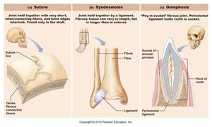
Fibrous Joints
Fibrous joints are connected by dense connective tissue and allow little to no movement. Types include:
Sutures: Found only in the skull; interlocking edges add strength.
Syndesmoses: Bones connected by ligaments (e.g., distal tibiofibular joint).
Gomphoses: Peg-in-socket joints (e.g., teeth in alveolar sockets).
Cartilaginous Joints
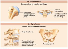
Cartilaginous joints unite bones with cartilage. They are classified as:
Synchondroses: Bones united by hyaline cartilage (e.g., epiphyseal plates, first rib and sternum).
Symphyses: Bones united by fibrocartilage (e.g., pubic symphysis, intervertebral discs).
Synovial Joints
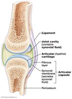
Synovial joints are the most common and movable type of joint in the body. They have a joint cavity filled with synovial fluid and are characterized by seven key features:
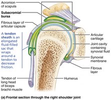
Articular cartilage: Cushions bone ends.
Joint (synovial) cavity: Space containing synovial fluid.
Articular capsule: Double-layered; outer fibrous layer and inner synovial membrane.
Synovial fluid: Lubricates and nourishes articular cartilage.
Reinforcing ligaments: Strengthen and stabilize the joint.
Nerves and blood vessels: Provide nutrients and monitor joint health.
High mobility: All synovial joints are diarthrotic (freely movable).
Additional Structures in Some Synovial Joints
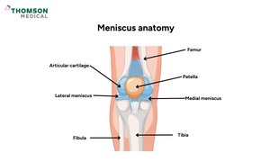
Fatty pads: Cushioning between capsule and bone.
Articular discs (menisci): Improve fit, stabilize, and reduce wear.
Bursae and Tendon Sheaths
Bursae: Fluid-filled sacs that reduce friction between moving structures.
Tendon sheaths: Elongated bursae that wrap around tendons to decrease friction.
Movements at Synovial Joints
Planes of Movement
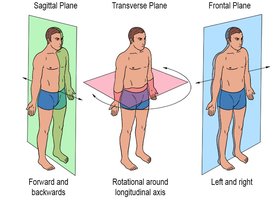
Movements at synovial joints are described relative to three anatomical planes:
Sagittal plane: Forward and backward movements.
Frontal (coronal) plane: Side-to-side movements.
Transverse plane: Rotational movements.
Types of Movements
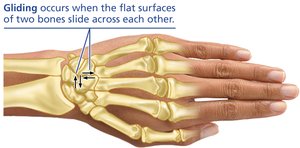
Gliding: Flat bone surfaces slide over each other (e.g., intercarpal joints).
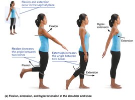
Angular movements: Change the angle between bones.
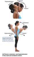
Flexion: Decreases the angle (e.g., bending the elbow).
Extension: Increases the angle (e.g., straightening the knee).
Hyperextension: Extension beyond anatomical position.
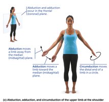
Abduction: Movement away from the midline.
Adduction: Movement toward the midline.
Circumduction: Limb describes a cone in space, combining flexion, abduction, extension, and adduction.
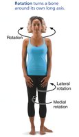
Rotation: Turning a bone around its own long axis.
Medial rotation: Toward the midline.
Lateral rotation: Away from the midline.
Special movements:
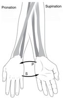
Supination and pronation: Rotation of the forearm (radius and ulna).
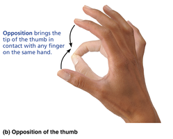
Opposition: Movement of the thumb to touch other fingers.
Major Synovial Joints: Structure and Movements
Shoulder Joint (Glenohumeral Joint)
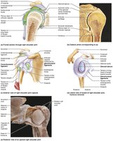
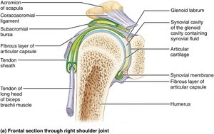
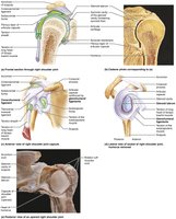
The shoulder joint is the most freely moving joint in the body, classified as a ball-and-socket synovial joint. Its structure allows for a wide range of motion but sacrifices stability.
Articular surfaces: Large head of humerus fits into shallow glenoid cavity of scapula.
Articular capsule: Thin and loose, enhancing mobility.
Reinforcing tendons: Tendon of long head of biceps brachii (superstabilizer) and four rotator cuff tendons encircle the joint.
Factors Influencing Joint Stability
Shape of articular surfaces: Shallow surfaces are less stable.
Ligament number and location: More ligaments increase strength.
Muscle tone: Most important factor; keeps tendons taut and stabilizes joints.
Clinical Relevance: Joint Injuries and Disorders
Common Joint Injuries
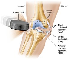
Cartilage tears: Often occur in menisci; poor repair due to low vascularization.
Sprains: Ligaments stretched or torn; common in ankle, knee, and back.
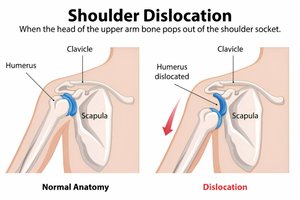
Dislocations: Bones forced out of alignment; shoulder is particularly susceptible due to its mobility.
Rheumatoid Arthritis (RA)
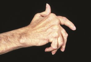
RA is a chronic, inflammatory, autoimmune disease where the immune system attacks joint tissues. It is more common in females and typically arises between ages 40 and 50. Symptoms include joint pain, swelling, anemia, osteoporosis, muscle weakness, and cardiovascular problems.
Summary Table: Movements at Synovial Joints
Movement | Description | Example |
|---|---|---|
Gliding | Flat surfaces slide over each other | Intercarpal joints |
Flexion | Decreases angle between bones | Bending elbow |
Extension | Increases angle between bones | Straightening knee |
Abduction | Movement away from midline | Raising arm laterally |
Adduction | Movement toward midline | Lowering arm to side |
Circumduction | Limb moves in a circle | Shoulder joint |
Rotation | Bone turns around its long axis | Turning head |
Supination/Pronation | Rotation of forearm | Turning palm up/down |
Opposition | Thumb touches other fingers | Grasping objects |
Key Points
Joints are classified by structure (fibrous, cartilaginous, synovial) and function (synarthroses, amphiarthroses, diarthroses).
Synovial joints are the most mobile and include features such as articular cartilage, synovial fluid, and reinforcing ligaments.
Movements at synovial joints include gliding, angular movements, rotation, and special movements.
The shoulder joint is highly mobile but prone to dislocation due to its structure.
Common joint injuries include sprains, cartilage tears, and dislocations; rheumatoid arthritis is a significant autoimmune joint disorder.
