 Indietro
Joints (Articulations): Structure, Classification, and Clinical Implications
Guida di studio - Note intelligenti
 Appunti personalizzati basati sui tuoi materiali, ampliati con definizioni chiave, esempi e contesto.
Joints (Articulations)
Introduction to Joints
Joints, also known as articulations, are the sites where two or more bones meet. They are essential for providing mobility to the skeleton and holding it together. Understanding joint structure and function is crucial for diagnosing and treating musculoskeletal injuries and diseases.
Joint Classifications
Functional Classification
Joints are classified based on the degree of movement they allow:
Synarthroses: Immovable joints
Amphiarthroses: Slightly movable joints
Diarthroses: Freely movable joints
Structural Classification
Structural classification is based on the material binding the bones and the presence or absence of a joint cavity:
Fibrous Joints: Bones joined by dense fibrous connective tissue; no joint cavity; mostly immovable.
Cartilaginous Joints: Bones united by cartilage; no joint cavity; not highly movable.
Synovial Joints: Bones separated by a fluid-filled joint cavity; all are diarthrotic (freely movable).
Types of Cartilaginous Joints
Cartilaginous joints are further divided into:
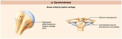
Synchondroses: Bones united by hyaline cartilage (e.g., epiphyseal plate, joint between first rib and sternum).
Symphyses: Bones united by fibrocartilage (e.g., intervertebral joints, pubic symphysis).
Synovial Joints
General Features of Synovial Joints
Synovial joints are characterized by the following features:
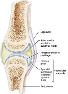
Articular cartilage: Hyaline cartilage covering bone ends, preventing crushing.
Joint (synovial) cavity: Small, fluid-filled space unique to synovial joints.
Articular capsule: Two layers—external fibrous layer (dense irregular connective tissue) and inner synovial membrane (loose connective tissue producing synovial fluid).
Reinforcing ligaments: Capsular, extracapsular, and intracapsular ligaments strengthen the joint.
Synovial fluid: Viscous, slippery filtrate of plasma and hyaluronic acid; lubricates and nourishes articular cartilage; contains phagocytic cells.
Nerves and blood vessels: Nerves detect pain and monitor joint position; capillary beds supply filtrate for synovial fluid.
Other Features of Synovial Joints
Fatty pads: Cushioning between fibrous layer and synovial membrane or bone.
Articular discs (menisci): Fibrocartilage improves fit, stabilizes joint, reduces wear.
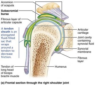
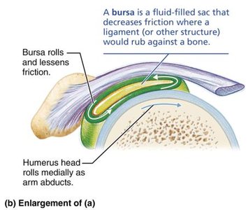
Bursae: Fluid-filled sacs reducing friction where ligaments, muscles, skin, tendons, or bones rub together.
Tendon sheaths: Elongated bursae wrapped around tendons subjected to friction.
Joint Stability
Factors Influencing Stability
Three main factors determine the stability of synovial joints:
Shape of articular surfaces: Deeper surfaces provide more stability (e.g., ball-and-socket joints).
Ligament number and location: More ligaments generally mean stronger joints.
Muscle tone: Most important factor; keeps tendons taut and reinforces joints, especially in the shoulder, knee, and foot arches.
Movements at Synovial Joints
Types of Movement
Synovial joints allow a variety of movements:
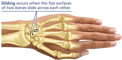
Gliding: Flat bone surfaces slide across each other (e.g., intercarpal joints).
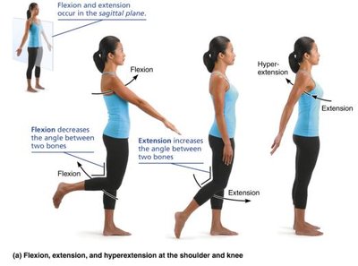
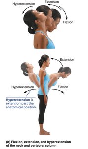
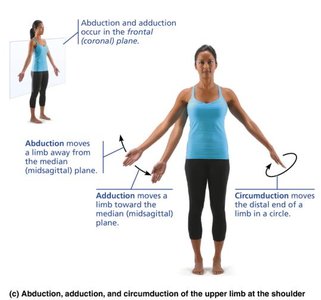
Angular movements: Change the angle between bones (flexion, extension, hyperextension, abduction, adduction, circumduction).
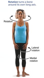
Rotation: Bone turns around its own long axis (medial and lateral rotation).
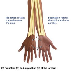
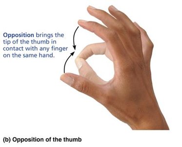
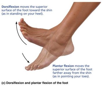
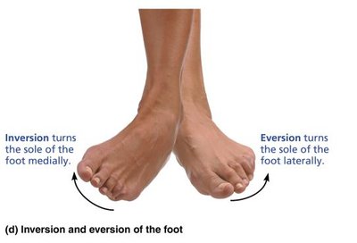
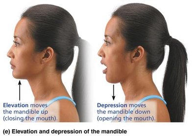
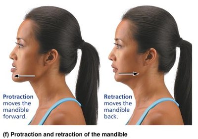
Special movements: Include supination/pronation, opposition, dorsiflexion/plantar flexion, inversion/eversion, elevation/depression, protraction/retraction.
Summary Table: Joint Movements
Movement | Definition |
|---|---|
Gliding | Sliding the flat surfaces of two bones across each other |
Flexion | Decreasing the angle between two bones, usually in the sagittal plane |
Extension | Increasing the angle between two bones, usually in the sagittal plane |
Abduction | Moving a limb away from the body midline in the frontal plane |
Adduction | Moving a limb toward the body midline in the frontal plane |
Circumduction | Moving a limb or finger so that it describes a cone in space |
Rotation | Turning a bone around its longitudinal axis |
Medial rotation | Rotating toward the median plane |
Lateral rotation | Rotating away from the median plane |
Types of Synovial Joints
Classification by Shape and Movement
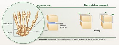
Synovial joints are classified into six types based on the shape of their articular surfaces and the movement they allow:
Plane: Nonaxial gliding movements (e.g., intercarpal joints).
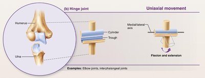
Hinge: Uniaxial movement (flexion and extension; e.g., elbow).
Pivot: Uniaxial rotation (e.g., proximal radioulnar joint).
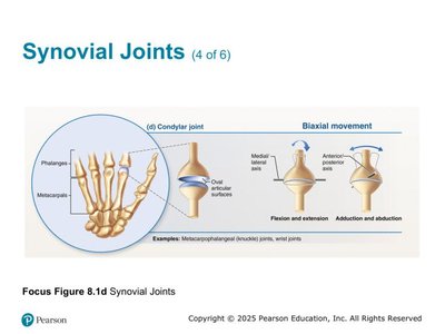
Condylar (Ellipsoid): Biaxial movement (flexion/extension, abduction/adduction; e.g., metacarpophalangeal joints).
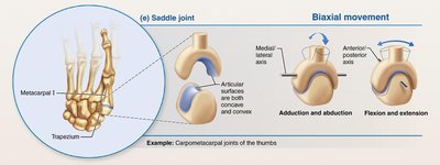
Saddle: Biaxial movement (e.g., carpometacarpal joint of thumb).
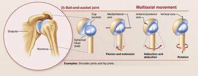
Ball-and-socket: Multiaxial movement (e.g., shoulder, hip).
Major Synovial Joints of the Body
Temporomandibular Joint (TMJ)
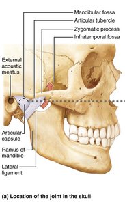
The TMJ is a unique synovial joint in the skull, prone to dislocation due to its shallow socket. Dislocation often occurs anteriorly, causing the mouth to remain open. Symptoms include pain, muscle tenderness, popping sounds, and stiffness. Treatment may involve realignment, bite plates, and muscle relaxation.
Shoulder Joint (Glenohumeral Joint)
The shoulder joint is the most freely moving joint in the body, classified as a ball-and-socket joint. Its stability is sacrificed for mobility, making it prone to dislocations, especially anteriorly and inferiorly. The glenoid cavity provides poor support when the humerus is rotated and abducted.
Elbow Joint
The elbow is a hinge joint formed primarily by the articulation between the humerus and ulna. It allows flexion and extension only, with ligaments restricting side-to-side movement.
Hip Joint (Coxal Joint)
The hip is a ball-and-socket joint with a deep socket (acetabulum) providing great stability. The large head of the femur articulates with the acetabulum, and reinforcing ligaments further stabilize the joint. Hip dislocations are rare due to this stability.
Knee Joint
The knee is the largest and most complex joint, consisting of three joints surrounded by a single cavity. It absorbs significant vertical force but is vulnerable to horizontal blows. Common injuries involve collateral ligaments, cruciate ligaments, and menisci, often requiring surgical repair.
Clinical Implications
Common Joint Injuries
Cartilage tears: Result from compression and shear stress; cartilage rarely repairs itself and may require arthroscopic surgery.
Sprains: Ligaments are stretched or torn; partial tears heal slowly, complete tears may require surgery or grafts.
Dislocations: Bones are forced out of alignment, often accompanied by sprains and inflammation; must be reduced to treat.
Bursitis: Inflammation of bursae, usually from blow or friction; treated with rest, ice, and anti-inflammatory drugs.
Tendonitis: Inflammation of tendon sheaths, typically from overuse; similar treatment to bursitis.
Arthritis
Arthritis encompasses over 100 types of inflammatory or degenerative joint diseases. Symptoms include pain, stiffness, and swelling. Acute forms are often bacterial and treated with antibiotics, while chronic forms include osteoarthritis, rheumatoid arthritis, and gouty arthritis.
Summary Table: Types of Synovial Joints
Type | Movement | Example |
|---|---|---|
Plane | Nonaxial (gliding) | Intercarpal joints |
Hinge | Uniaxial (flexion/extension) | Elbow |
Pivot | Uniaxial (rotation) | Proximal radioulnar joint |
Condylar | Biaxial (flexion/extension, abduction/adduction) | Metacarpophalangeal joints |
Saddle | Biaxial (flexion/extension, abduction/adduction) | Thumb joint |
Ball-and-socket | Multiaxial (all movements) | Shoulder, hip |
Key Terms: Articulation, synovial fluid, ligament, cartilage, bursae, tendon sheath, flexion, extension, abduction, adduction, rotation, arthritis, sprain, dislocation.
