 Indietro
Joints: Structure, Classification, and Function
Guida di studio - Note intelligenti
 Appunti personalizzati basati sui tuoi materiali, ampliati con definizioni chiave, esempi e contesto.
Joints (Articulations)
Definition and Overview
Joints, also known as articulations or arthroses, are the sites where two or more skeletal components meet. They may allow movement or be immobile, depending on their structure and function. Joints are essential for providing mobility, stability, and support to the skeletal system.
Classification of Joints
Joints are classified in two main ways: structurally and functionally.
Structural Classification: Based on the material binding the bones and the presence or absence of a joint cavity.
Fibrous joints
Cartilaginous joints
Synovial joints
Functional Classification: Based on the degree of movement permitted.
Synarthrosis: Immobile
Amphiarthrosis: Slightly movable
Diarthrosis: Freely movable
Fibrous Joints
General Structure
Fibrous joints are characterized by the absence of a synovial cavity and the presence of dense irregular connective tissue binding the bones. These joints are generally immobile or allow very limited movement.
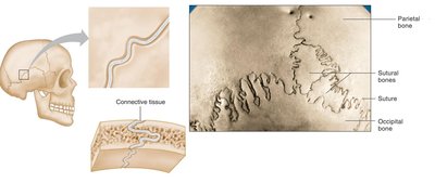
Suture: Found only in the skull, sutures are joints held together by very short, interconnecting fibers. The bone edges interlock, providing stability.
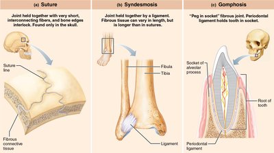
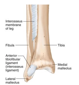
Syndesmosis: Bones are united by a ligament or interosseous membrane, allowing more movement than sutures. Example: Inferior tibiofibular joint.
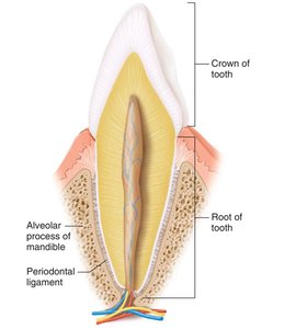
Gomphosis: A peg-in-socket joint where the root of a tooth fits into the socket of the jaw, held by fibrous tissue (periodontal ligament).
Cartilaginous Joints
General Structure
Cartilaginous joints lack a synovial cavity and are held together by cartilage. They are classified as primary or secondary cartilaginous joints.
Primary Cartilaginous Joint (Synchondrosis): Bones are united by hyaline cartilage, typically immobile and temporary. Example: Epiphyseal plates in growing bones.
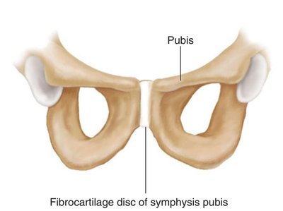
Secondary Cartilaginous Joint (Symphysis): Bones are united by fibrocartilage and hyaline cartilage, usually located in the midline, permanent, and slightly movable. Example: Pubic symphysis.
Synovial Joints
General Structure
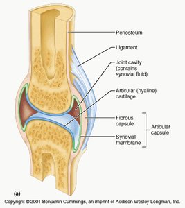
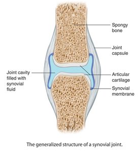
Synovial joints are the most movable type of joint and are characterized by the presence of a synovial cavity. The bones are held together by dense irregular connective tissue and are surrounded by an articular capsule.
Articular hyaline cartilage: Covers the ends of bones, reducing friction and absorbing shock.
Synovial cavity: Space between the bones filled with synovial fluid.
Synovial fluid: Lubricates the joint and nourishes the cartilage.
Articular capsule: Encloses the joint cavity, consisting of a fibrous layer and a synovial membrane.
Ligaments: Strengthen and stabilize the joint.
Intra-articular structures: Include cartilages, ligaments, and muscles within the joint.
Classification of Synovial Joints by Shape and Movement
Synovial joints are classified according to the shape of their articular surfaces and the type of movement they allow:
Plane Joint: Flat or slightly curved surfaces; allows gliding movements. Example: Intercarpal joints.
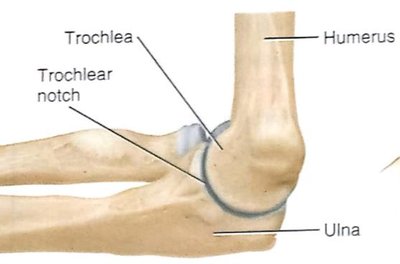
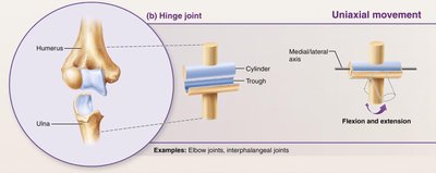
Hinge Joint: Convex surface fits into concave surface; uniaxial movement (flexion and extension). Example: Elbow joint.
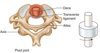
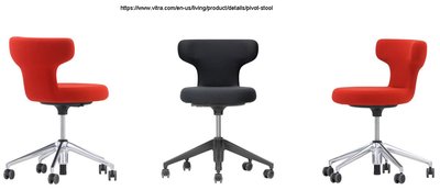
Pivot Joint: Rounded or pointed surface articulates with a ring; uniaxial rotatory movement. Example: Atlantoaxial joint.
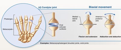
Ellipsoid (Condylar) Joint: Oval-shaped projection fits into oval depression; biaxial movement (flexion-extension, abduction-adduction). Example: Metacarpophalangeal joints.
Saddle Joint: One bone is saddle-shaped, the other fits like a rider; biaxial movement. Example: Thumb carpometacarpal joint.
Ball & Socket Joint: Ball-like surface fits into a cuplike depression; multiaxial movement. Example: Shoulder and hip joints.

Joint Movements
Types of Movement
Gliding: Occurs in plane joints; one bone slips over another.
Angular: Change in angle between bones.
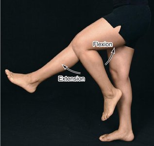
Flexion and extension: Occur around transverse axis.
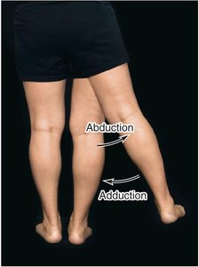
Abduction and adduction: Occur around anteroposterior axis.
Rotation: Movement around vertical/longitudinal axis. Example: Circumduction at shoulder and hip joints.
Special Movements: Include elevation, depression, protraction, retraction, supination, pronation, inversion, eversion, dorsiflexion, and plantar flexion.
Factors Limiting Range of Movement
Shape of articulating bones: Disproportionate surfaces affect movement freedom.
Tension of ligaments: Ligament tension restricts movement.
Tension of antagonistic muscles: Muscle tension can arrest movement.
Approximation of soft parts: Soft tissue contact limits movement.
Hormones: Hormones like relaxin increase flexibility, especially in the pelvis during childbirth.
Factors Maintaining Stability of Joints
Bony configuration
Ligaments
Muscles
Atmospheric pressure
Bursae and Tendon Sheaths
Bursae
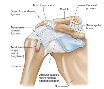
Bursae are closed fibrous sacs lined with a smooth membrane and filled with viscous fluid. They reduce friction where tendons rub against bones, ligaments, or other tendons, commonly found near joints.
Synovial Sheath
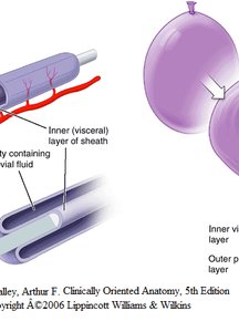
A synovial sheath is a tubular bursa that surrounds a tendon, reducing friction as the tendon passes under ligaments or through osseofibrous tunnels. The mesotendon allows blood vessels to enter the tendon.
Aging and Joints
Effects of Aging
Decreased production of synovial fluid
Thinning of articular cartilage
Shortening and loss of flexibility in ligaments
Degenerative changes may begin as early as age 20, with most people affected by their 80s
Knee, hip, elbow, shoulder, and vertebral column are commonly affected
Common Joint Injuries and Disorders
Arthritis
Arthritis is a group of disorders characterized by inflammation of joints, leading to pain, stiffness, and reduced mobility. Common types include osteoarthritis, rheumatoid arthritis, and gout.
Lyme Disease
Lyme disease is caused by the bacterium Borrelia burgdorferi, transmitted by ticks. It can lead to joint pain, swelling, and long-term complications if untreated.
References
Hole’s Anatomy and Physiology, 12th edition
Gray’s Anatomy for Students
Tortora’s Principles of Anatomy & Physiology – Global edition
