 Indietro
Joints: Structure, Classification, and Function in Human Anatomy
Guida di studio - Note intelligenti
 Appunti personalizzati basati sui tuoi materiali, ampliati con definizioni chiave, esempi e contesto.
Joints and Their Classification
Definition and Functions of Joints
Joints, also known as articulations, are points where two bones meet. They play crucial roles in the human body by enabling movement, providing stability, and allowing long bones to lengthen during growth. The scientific study of joints is called arthrology.
Enable movement: Joints allow bones to move when muscles and tendons exert force across them.
Provide stability: Some joints restrict movement to protect underlying structures.
Allow bone growth: The epiphyseal plate is a temporary joint where long bones grow in length during development.
Joint names are typically derived from the bones involved (e.g., atlantooccipital joint, radioulnar joint).
Classification of Joints
Joints are classified by their freedom of movement and by how adjacent bones are joined:
Diarthrosis: Freely movable joints
Amphiarthrosis: Slightly movable joints
Synarthrosis: Little or no movement
Structurally, joints are classified as fibrous, cartilaginous, bony, or synovial.
Fibrous and Cartilaginous Joints
Fibrous Joints (Synarthrosis)
Fibrous joints are points where adjacent bones are bound by collagen fibers. They are generally immovable and include three types:
Sutures: Immovable joints that bind skull bones together. Occur only in the skull.
Gomphoses: Attachments of teeth to their sockets, held by the periodontal ligament.
Syndesmoses: Two bones bound by longer collagenous fibers, allowing more mobility (e.g., interosseus membrane between radius and ulna).
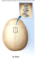
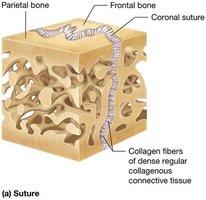
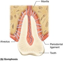
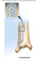
Cartilaginous Joints
Cartilaginous joints are where two bones are linked by cartilage. There are two types:
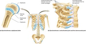
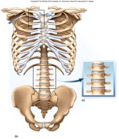
Synchondroses: Bones joined by hyaline cartilage (e.g., first sternocostal and costochondral joints, epiphyseal plate in children).
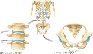
Symphyses: Bones joined by fibrocartilage (e.g., pubic symphysis, intervertebral discs). Allow slight movement.
Synovial Joints (Diarthroses)
Anatomical Components of Synovial Joints
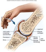
Synovial joints are characterized by a joint cavity separating two bones. Most are freely movable and have several key anatomical features:
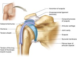
Articular capsule: Encloses the joint cavity, continuous with periosteum, lined by synovial membrane.
Synovial fluid: Slippery fluid that lubricates, nourishes cartilage, and absorbs shock.
Articular cartilage: Hyaline cartilage covering joint surfaces.
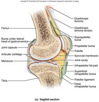
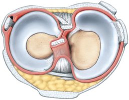
Articular discs and meniscus: Fibrocartilage pads that absorb shock, guide bone movements, and stabilize joints.
Tendons: Attach muscle to bone, providing stabilization.
Ligaments: Attach bone to bone, supporting and reinforcing joints.
Functions of Synovial Fluid
Lubrication: Reduces friction between articular cartilage layers.
Nutrient distribution: Circulates during movement, providing nutrients to chondrocytes.
Shock absorption: Cushions joints by distributing shocks across articular surfaces.
Exercise and Articular Cartilage
Exercise warms synovial fluid, making it less viscous and more easily absorbed by cartilage. This swelling provides a more effective cushion against compression. Regular exercise is essential for cartilage health, as it promotes nutrient and waste exchange.
Articular Discs and Menisci
Some synovial joints contain fibrocartilage structures called articular discs and menisci. These absorb shock, guide bone movements, improve fit between bones, and stabilize joints.
Tendons and Ligaments
Synovial joints allow more mobility but less stability than other joint types. Tendons and ligaments provide additional stabilization:
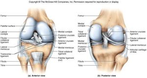
Tendon: Tough collagenous tissue attaching muscle to bone; stabilizes joints during muscle contraction.
Ligament: Similar tissue attaching bone to bone; supports, strengthens, and reinforces synovial joints.
Tendon Sheaths and Bursae
Bursae and tendon sheaths are synovial fluid-filled structures that minimize friction and provide stabilization in high-stress regions:
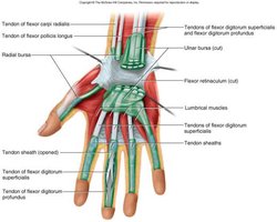
Bursa: Saclike extension of joint capsule between structures to reduce friction.
Tendon sheath: Cylinder of connective tissue lined with synovial membrane, wrapped around a tendon.
Joint Disorders
Bursitis
Bursitis is inflammation of a bursa, often caused by trauma, repetitive movements, or inflammatory disease. Common sites include the shoulder, elbow, hip, and knee. Treatment focuses on reducing pain and swelling.
Arthritis
Arthritis is inflammation of one or more joints, resulting in pain and limited movement. Common types include:
Osteoarthritis: Most common; caused by wear and tear, injuries, and aging.
Rheumatoid arthritis: Autoimmune attack on joint tissues.
Gouty arthritis: Inflammatory reaction to uric acid crystal deposits.
Arthroplasty is the replacement of a diseased joint with an artificial device (prosthesis).
Function and Types of Synovial Joints
Functional Classes of Synovial Joints
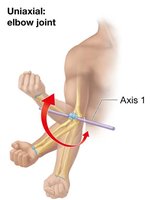
Synovial joints are classified by the number of axes around which bones move:
Nonaxial: Motion in one or more planes without moving around an axis.
Uniaxial: Motion around one axis (e.g., elbow joint).
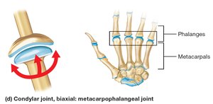
Biaxial: Motion around two axes (e.g., metacarpophalangeal joints).
Multiaxial (triaxial): Motion around three axes (e.g., shoulder joint).
Movements of Synovial Joints
Joint movements are described relative to the anatomical position (zero position). Common movements include:
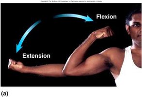
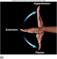
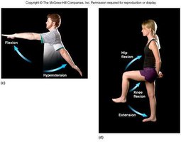
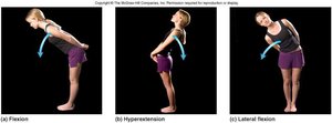
Flexion: Decreases the angle of a joint.
Extension: Straightens and returns to anatomical position.
Hyperextension: Extension beyond 180 degrees.
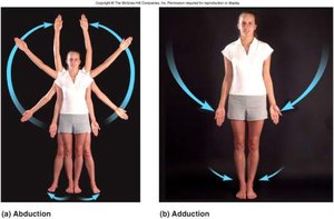
Abduction: Movement away from the midline.
Adduction: Movement toward the midline.
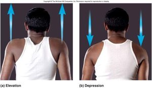
Elevation: Raises a bone vertically.
Depression: Lowers a bone vertically.
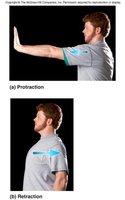
Protraction: Moves a part anteriorly.
Retraction: Moves a part posteriorly.
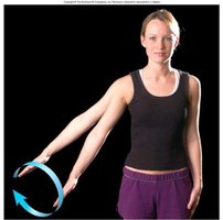
Circumduction: Circular movement of an appendage.
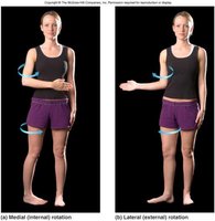
Rotation: Movement on a longitudinal axis (medial or lateral).
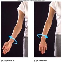
Supination: Rotation of forearm so palm faces forward.
Pronation: Rotation of forearm so palm faces rearward.
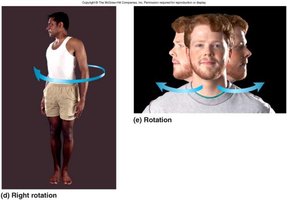
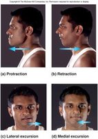
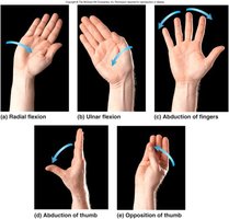
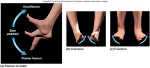
Types of Synovial Joints
There are six structural types of synovial joints, each allowing specific movements:
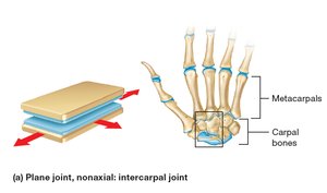
Plane (gliding) joint: Articulation between flat surfaces; least mobile.
Hinge joint: Convex surface interacts with concave depression; uniaxial movement.
Pivot joint: Rounded end fits into groove; uniaxial rotation.
Condylar (ellipsoid) joint: Oval, convex surface fits into shallow concave surface; biaxial movement.
Saddle joint: Each surface has concave and convex regions; biaxial movement.
Ball-and-socket joint: Spherical surface fits into cup-shaped depression; multiaxial movement.
Stability vs. Mobility
There is a trade-off between stability and mobility in joints. The most mobile joints are the most easily injured.
Comparative Anatomy of Major Joints
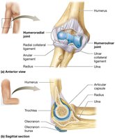
Elbow: Stable hinge joint with two articulations and three strong ligaments (humeroulnar and humeroradial joints).
Knee: Tibiofemoral and patellofemoral joints; medial and lateral meniscus; susceptible to injury, especially in contact sports.
Shoulder: Ball-and-socket joint; highly mobile but less stable; supported by rotator cuff muscles and tendons.
Hip: Very stable multiaxial joint; acetabular labrum stabilizes the head of the femur.
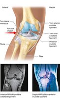
Example: The knee joint is stabilized by menisci and ligaments but is vulnerable to injury from lateral blows, which can result in the "unhappy triad" (damage to tibial collateral ligament, medial meniscus, and anterior cruciate ligament).
Additional info: The notes above expand on brief points from the original material, providing definitions, examples, and academic context for each joint type and movement.
