 Indietro
Joints: Structure, Classification, and Movements
Guida di studio - Note intelligenti
 Appunti personalizzati basati sui tuoi materiali, ampliati con definizioni chiave, esempi e contesto.
Joints (Articulations)
Introduction to Joints
Joints, also known as articulations, are the sites where two or more bones meet. They serve two primary functions: providing mobility to the skeleton and holding the skeleton together. Understanding the nature of joints is essential for diagnosing and treating injuries such as ankle sprains.
Classification of Joints
Structural Classification: Based on the material binding the bones and the presence or absence of a joint cavity. The three types are:
Fibrous Joints
Cartilaginous Joints
Synovial Joints
Functional Classification: Based on the degree of movement allowed. The three types are:
Synarthroses: Immovable joints
Amphiarthroses: Slightly movable joints
Diarthroses: Freely movable joints
Fibrous Joints
General Features
Fibrous joints are characterized by bones joined by dense fibrous connective tissue, with no joint cavity. Most are immovable, though the degree of movement depends on the length of connective tissue fibers. There are three types:
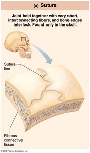
Sutures: Rigid, interlocking joints found only in the skull. They allow for growth during youth and ossify in middle age, forming immovable joints called synostoses.
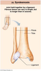
Syndesmoses: Bones connected by ligaments. The length of the fibers determines the amount of movement. Short fibers (e.g., inferior tibiofibular joint) allow little movement, while longer fibers (e.g., interosseous membrane between radius and ulna) allow more movement.
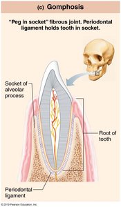
Gomphoses: Peg-in-socket joints, such as the teeth in alveolar sockets. The fibrous connection is the periodontal ligament.
Cartilaginous Joints
General Features
Cartilaginous joints are formed by bones united by cartilage, with no joint cavity. They are not highly movable. There are two main types:
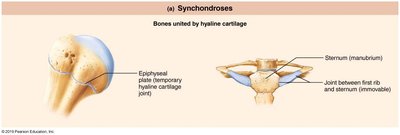
Synchondroses: Bones united by a bar or plate of hyaline cartilage. Most are synarthrotic (immovable). Examples include the temporary epiphyseal plate joints and the joint between the first rib and the manubrium of the sternum.
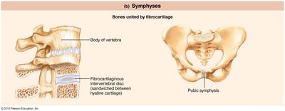
Symphyses: Bones united by fibrocartilage, with hyaline cartilage present as articular cartilage on bony surfaces. Symphyses are strong, amphiarthrotic (slightly movable) joints. Examples include intervertebral joints and the pubic symphysis.
Synovial Joints
General Features
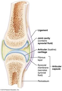
Synovial joints are characterized by bones separated by a fluid-filled joint cavity. All synovial joints are diarthrotic (freely movable) and include almost all limb joints. They have six general features:
Articular cartilage: Hyaline cartilage covering bone ends, preventing crushing.
Joint (synovial) cavity: Small, fluid-filled space unique to synovial joints.
Articular (joint) capsule: Two layers: external fibrous layer (dense irregular connective tissue) and inner synovial membrane (loose connective tissue producing synovial fluid).
Synovial fluid: Viscous, slippery filtrate of plasma and hyaluronic acid, lubricating and nourishing articular cartilage. Contains phagocytic cells to remove debris.
Reinforcing ligaments: Capsular (thickened part of fibrous layer), extracapsular (outside capsule), and intracapsular (deep to capsule, covered by synovial membrane).
Nerves and blood vessels: Nerves detect pain and monitor joint position; capillary beds supply filtrate for synovial fluid.
Other Features
Fatty pads: Cushioning between fibrous layer and synovial membrane or bone.
Articular discs (menisci): Fibrocartilage separates articular surfaces to improve fit, stabilize joint, and reduce wear and tear.
Bursae and Tendon Sheaths
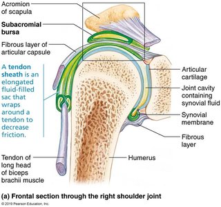
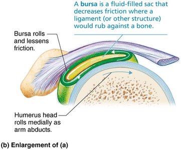
Bursae are fluid-filled sacs that reduce friction where ligaments, muscles, skin, tendons, or bones rub together. Tendon sheaths are elongated bursae wrapped around tendons subjected to friction.
Factors Influencing Stability
Shape of articular surface: Shallow surfaces are less stable than ball-and-socket joints.
Ligament number and location: More ligaments provide greater strength.
Muscle tone: Keeps tendons taut as they cross joints, crucial for reinforcing shoulder, knee, and foot arches.
Movements Allowed by Synovial Joints
General Principles
All muscles attach to bone or connective tissue at no fewer than two points: origin (immovable bone) and insertion (movable bone). Muscle contraction causes insertion to move toward origin. Movements occur along transverse, frontal, or sagittal planes.
Types of Movement
Gliding: One flat bone surface glides over another. Examples: intercarpal, intertarsal, and vertebral articular process joints.
Angular Movements: Increase or decrease angle between bones.
Flexion: Decreases joint angle.
Extension: Increases joint angle.
Hyperextension: Movement beyond anatomical position.
Abduction: Movement away from midline.
Adduction: Movement toward midline.
Circumduction: Limb describes a cone in space, combining flexion, abduction, extension, and adduction.
Rotation: Turning bone around its own long axis.
Medial rotation: Toward midline.
Lateral rotation: Away from midline.
Examples: rotation between C1 and C2 vertebrae, rotation of humerus and femur.
Special Movements:
Supination and pronation: Rotation of radius and ulna.
Dorsiflexion and plantar flexion: Movements of the foot.
Inversion and eversion: Movements of the sole of the foot.
Protraction and retraction: Movements of the mandible.
Elevation and depression: Lifting or lowering body parts.
Opposition: Movement of thumb to touch other fingers.
Types of Synovial Joints
Classification by Shape and Movement
Synovial joints are classified into six types based on the shape of their articular surfaces and the movements they allow:
Plane
Hinge
Pivot
Condylar
Saddle
Ball-and-socket
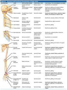
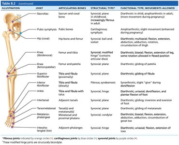
Structural and Functional Characteristics of Body Joints
Comparison Table
The following table summarizes the structural and functional characteristics of major body joints, including their articulating bones, structural type, and functional type/movements allowed.
Joint | Articulating Bones | Structural Type | Functional Type / Movements Allowed |
|---|---|---|---|
Skull | Cranial bones | Fibrous, suture | Synarthrotic, no movement |
Inferior tibiofibular | Tibia and fibula | Fibrous, syndesmosis | Synarthrotic, slight movement |
Tooth in socket | Tooth and alveolar socket | Fibrous, gomphosis | Synarthrotic, no movement |
Epiphyseal plate | Epiphysis and diaphysis | Cartilaginous, synchondrosis | Synarthrotic, no movement |
Intervertebral | Vertebrae | Cartilaginous, symphysis | Amphiarthrotic, slight movement |
Shoulder | Scapula and humerus | Synovial, ball-and-socket | Diarthrotic, multiaxial movement |
Elbow | Humerus and ulna | Synovial, hinge | Diarthrotic, uniaxial movement |
Hip | Hip bone and femur | Synovial, ball-and-socket | Diarthrotic, multiaxial movement |
Knee | Femur and tibia | Synovial, modified hinge | Diarthrotic, biaxial movement |
Summary
Joints are essential for skeletal mobility and stability. Their classification, structure, and function are fundamental concepts in anatomy and physiology, with direct clinical relevance for understanding movement and treating joint injuries.
