 Indietro
Joints: Structure, Function, and Clinical Relevance
Guida di studio - Note intelligenti
 Appunti personalizzati basati sui tuoi materiali, ampliati con definizioni chiave, esempi e contesto.
Joints and Their Classification
Overview of Joint Types
Joints, also known as articulations, are regions where two or more bones meet. They are classified based on their structure and the range of motion they allow.
Fibrous Joints: Immovable joints held together by dense connective tissue.
Cartilaginous Joints: Slightly movable joints connected by cartilage.
Synovial Joints: Freely movable joints surrounded by a joint capsule and containing synovial fluid.
Intervertebral Joints
Structure and Function
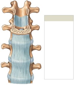
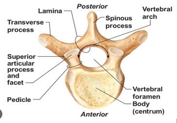
Intervertebral joints are located between the vertebrae of the spinal column. They allow for a range of movements and provide stability to the spine.
Movements: Flexion, extension, lateral flexion, and rotation.
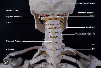
Atlantooccipital Joint: Synovial joint between the atlas (C1) and the occipital bone, allowing nodding motion.
Atlas and Axis: The first two cervical vertebrae (C1 and C2) are joined by a synovial joint, enabling rotation of the head.
Intervertebral Discs: Symphysis joints between vertebral bodies, composed of fibrocartilage.
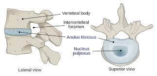
Intervertebral Disc Anatomy
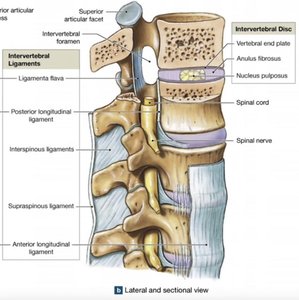
The intervertebral disc separates vertebral bodies and absorbs shock.
Anulus Fibrosus: Tough outer layer of fibrocartilage that attaches the disc to vertebrae.
Nucleus Pulposus: Elastic, gelatinous core that absorbs shocks.
Vertebral End Plates: Cartilage covering the superior and inferior surfaces of the disc.
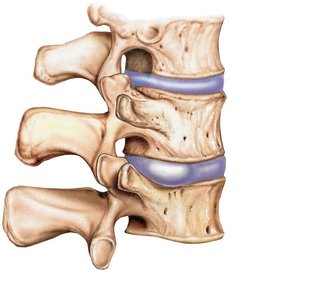
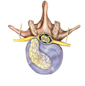
Disc Pathology
Damage to intervertebral discs can lead to clinical conditions:
Bulging Disc: The anulus fibrosus bulges and may invade the vertebral canal.
Herniated Disc: The nucleus pulposus breaks through the anulus fibrosus, compressing spinal nerves.
Intervertebral Ligaments
Ligaments stabilize the vertebral column and bind vertebrae together.
Ligamenta Flava: Connects the laminae of adjacent vertebrae.
Supraspinous Ligament: Attaches to the tips of spinous processes from C7 to the sacrum.
Interspinous Ligament: Connects the spinous processes of adjacent vertebrae.
Posterior Longitudinal Ligament: Connects the posterior surfaces of vertebral bodies.
Anterior Longitudinal Ligament: Connects the anterior surfaces of vertebral bodies.
Elbow Joint
Structure and Articulations
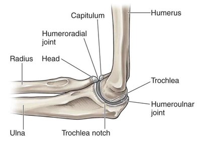
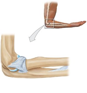
The elbow is a classic example of a hinge joint, allowing flexion and extension.
Humeroulnar Joint: Largest and strongest, between the trochlea of the humerus and the trochlear notch of the ulna.
Humeroradial Joint: Smaller, between the capitulum of the humerus and the head of the radius.
Supporting Structures
Biceps Brachii Muscle: Attaches to the radial tuberosity and controls elbow motion.
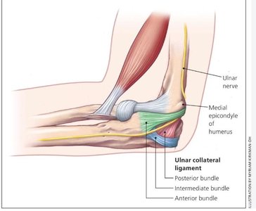
Elbow Ligaments: Radial collateral, annular, and ulnar collateral ligaments stabilize the joint.
Knee Joint
Structure and Function
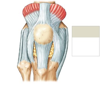
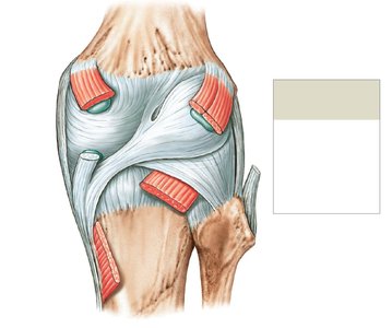
The knee is a complex hinge joint that transfers weight from the femur to the tibia.
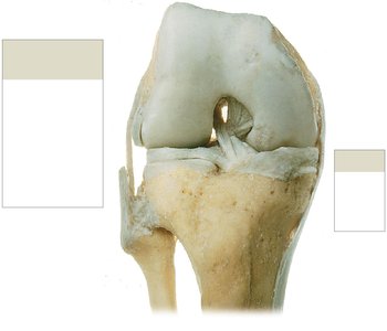
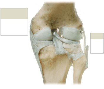
Articulations: Two femur-tibia articulations (medial and lateral condyles) and one between the patella and patellar surface of the femur.
Menisci: Fibrocartilage pads that cushion and stabilize the joint.
Major Supporting Ligaments
Patellar Ligament: Anterior support.
Popliteal Ligaments: Posterior support.
Anterior Cruciate Ligament (ACL): Inside joint capsule, prevents anterior displacement of tibia.
Posterior Cruciate Ligament (PCL): Inside joint capsule, prevents posterior displacement of tibia.
Tibial Collateral Ligament: Medial support.
Fibular Collateral Ligament: Lateral support.
Shoulder Joint
Structure and Supporting Elements
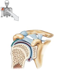
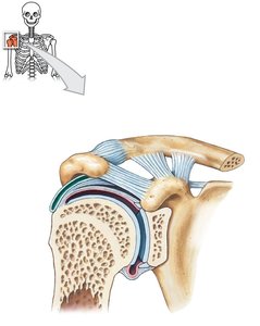
The shoulder (glenohumeral) joint is a ball-and-socket diarthrosis, allowing a wide range of motion.
Articulation: Between the head of the humerus and the glenoid cavity of the scapula.
Glenoid Labrum: Rim of fibrocartilage that deepens the socket.
Acromion and Coracoid Process: Project laterally and help stabilize the joint.
Shoulder Ligaments
Acromioclavicular Ligament
Coracoclavicular Ligament
Coraco-acromial Ligament
Coracohumeral Ligament
Glenohumeral Ligament
Rotator Cuff Muscles
Supraspinatus
Infraspinatus
Teres Minor
Subscapularis
Hip Joint
Structure and Function
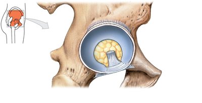
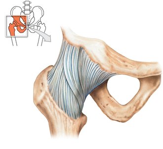
The hip joint is a strong ball-and-socket diarthrosis between the head of the femur and the acetabulum of the hip bone.
Acetabular Labrum: Rim of fibrocartilage that increases depth and seals in synovial fluid.
Ligaments: Iliofemoral, pubofemoral, ischiofemoral, transverse acetabular, and ligament of the femoral head.
Effects of Aging on Joints
Degenerative Changes and Diseases
With advancing age, joint mobility is often impaired due to degenerative changes.
Rheumatism: Pain and stiffness in the musculoskeletal system.
Arthritis: Inflammation of joints, including osteoarthritis (wear and tear), rheumatoid arthritis (autoimmune), and gouty arthritis (uric acid crystals).
Degenerative Changes: Immobilization reduces synovial fluid flow, increasing arthritis symptoms.
Bone Mass: Decreases with age, increasing fracture risk.
Integration with Other Systems
Skeletal System Functions and Interactions
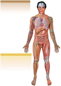
The skeletal system supports and stores energy and minerals for other body systems.
Bone Remodeling: Involves osteoblasts (formation) and osteoclasts (recycling).
Factors Affecting Bone Balance: Age, physical stress, hormone levels, calcium and phosphorus uptake, genetic/environmental factors.
Interactions: Muscles attach to bones, endocrine system controls bone growth, digestive and urinary systems provide minerals.
Summary Table: Major Joint Types and Their Features
Joint Type | Structure | Movement | Example |
|---|---|---|---|
Fibrous | Dense connective tissue | Immovable | Sutures of skull |
Cartilaginous | Cartilage | Slightly movable | Intervertebral discs |
Synovial | Joint capsule, synovial fluid | Freely movable | Shoulder, hip, knee, elbow |
Key Equations
Joint Stability:
Bone Remodeling:
Example: Clinical Application
Herniated Disc: Compression of spinal nerves can cause pain, numbness, or weakness in limbs.
ACL Injury: Common in athletes, leading to knee instability.
Additional info: Academic context was added to clarify joint types, clinical relevance, and integration with other systems.
