 Indietro
Lab 1: Language of Anatomy and Microscope Study Guide
Guida di studio - Note intelligenti
 Appunti personalizzati basati sui tuoi materiali, ampliati con definizioni chiave, esempi e contesto.
Language of Anatomy
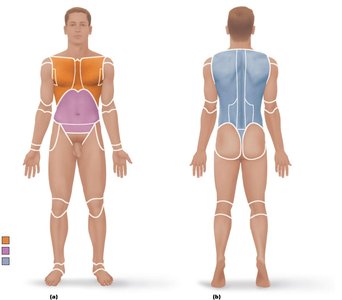
Anatomical Position
The anatomical position is a standardized visual reference point used in anatomy to ensure consistency when describing locations and directions on the human body. In this position, the person stands erect with feet together, eyes forward, and palms facing anteriorly with thumbs pointed away from the body.
Purpose: Provides a universal frame of reference for anatomical terminology.
Application: All directional terms are based on the body being in this position.
Directional Terminology
Directional terms are used to describe the locations of structures relative to other structures or locations in the body. These terms are essential for clear communication in anatomy.
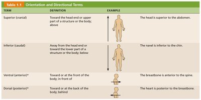
Superior (cranial): Toward the head end or upper part of a structure; above.
Inferior (caudal): Away from the head end or toward the lower part of a structure; below.
Ventral (anterior): Toward or at the front of the body; in front of.
Dorsal (posterior): Toward or at the back of the body; behind.
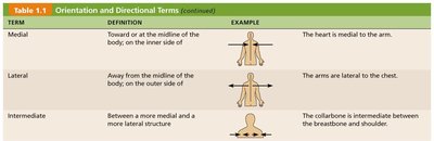
Medial: Toward or at the midline of the body; on the inner side.
Lateral: Away from the midline of the body; on the outer side.
Intermediate: Between a more medial and a more lateral structure.
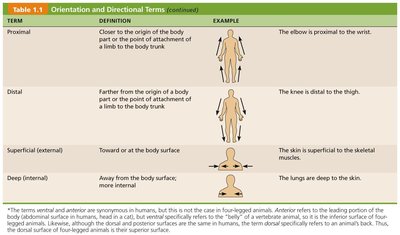
Proximal: Closer to the origin of a body part or the point of attachment of a limb to the body trunk.
Distal: Farther from the origin of a body part or the point of attachment of a limb to the body trunk.
Superficial (external): Toward or at the body surface.
Deep (internal): Away from the body surface; more internal.
Examples: The heart is superior to the abdomen; the navel is inferior to the chin; the breastbone is anterior to the spine; the heart is posterior to the breastbone; the heart is medial to the arm; the arms are lateral to the chest; the collarbone is intermediate between the breastbone and shoulder; the elbow is proximal to the wrist; the knee is distal to the thigh; the skin is superficial to the skeletal muscles; the lungs are deep to the skin.
Body Planes and Sections
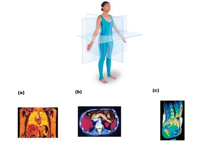
Body planes are imaginary lines that divide the body into sections, which are useful for anatomical study and medical imaging.
Frontal (coronal) plane: Divides the body into anterior and posterior parts.
Transverse (horizontal) plane: Divides the body into superior and inferior parts.
Median (midsagittal) plane: Divides the body into left and right halves.
Application: These planes are used in medical imaging (e.g., MRI scans) to view internal structures.
Body Cavities
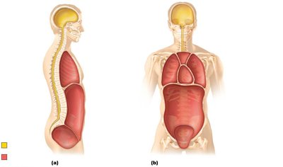
The human body contains two major sets of internal cavities: dorsal and ventral. These cavities house and protect vital organs.
Dorsal body cavity: Includes the cranial cavity (contains the brain) and vertebral cavity (contains the spinal cord).
Ventral body cavity: Includes the thoracic cavity (contains heart and lungs) and abdominopelvic cavity (contains digestive organs, urinary bladder, reproductive organs, and rectum).
The Microscope
Microscope Parts and Functions
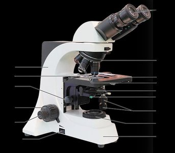
The compound light microscope is an essential tool in anatomy and physiology for observing cells and tissues. Understanding its parts and their functions is crucial for proper use.
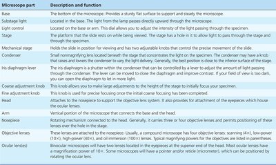
Base: Provides a sturdy flat surface to support and steady the microscope.
Substage light: Light source located in the base.
Light control: Adjusts the intensity of the light passing through the specimen.
Stage: Platform where slides are placed for viewing.
Mechanical stage: Holds the slide in position and allows precise movement.
Condenser: Focuses light on the specimen.
Iris diaphragm lever: Adjusts the amount of light passing through the specimen.
Coarse adjustment knob: Moves the stage up and down for initial focusing.
Fine adjustment knob: Allows for precise focusing once coarse focus is achieved.
Head: Supports the ocular lenses.
Nosepiece: Rotates to change objective lenses.
Objective lenses: Provide different levels of magnification (e.g., 4X, 10X, 40X, 100X).
Ocular lenses: Eyepieces, typically 10X magnification.
Arm: Supports the head and attaches to the base.
Total Magnification and Working Distance
Total magnification is calculated by multiplying the magnification of the objective lens by the magnification of the ocular lens. Working distance is the space between the bottom of the objective lens and the slide when the specimen is in focus.
Formula:
Working distance: Decreases as objective lens magnification increases.
Preparing a Wet Mount
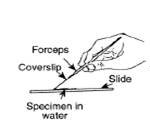
A wet mount is a technique used to prepare specimens for microscopic examination. The specimen is placed in water on a slide and covered with a cover slip.
Cut out the specimen (e.g., letter "e") and place it on the glass slide.
Cover with a clean cover slip.
Add a drop of water at the edge of the cover slip to allow water to be drawn under the slide.
Measuring Field Diameter
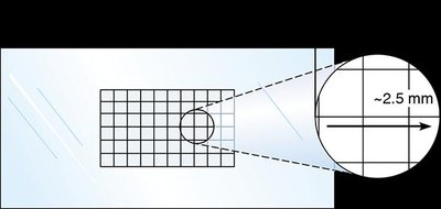
Field diameter is the width of the visible area under the microscope. It can be measured using a grid or graph paper.
Place graph paper under the slide and count the number of boxes across the field of view.
Field diameter decreases as magnification increases.
Estimate partial boxes if necessary.
Summary Chart for Microscope
The summary chart helps record and compare the properties of different objective lenses, including magnification, working distance, detail observed, and field diameter.
Lens | Magnification | Working Distance | Field Diameter |
|---|---|---|---|
Scanning (4X) | 4X × 10X = 40X | Largest | Largest |
Low Power (10X) | 10X × 10X = 100X | Medium | Medium |
High Power (40X) | 40X × 10X = 400X | Smallest | Smallest |
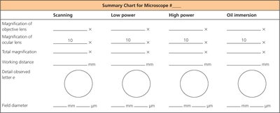
Example: At higher magnification, the field diameter and working distance decrease, allowing for more detailed observation of the specimen.
Additional info:
Microscope use is fundamental in anatomy for studying cells, tissues, and microorganisms.
Understanding anatomical terminology and microscope operation is essential for success in laboratory and clinical settings.
