 Indietro
Lymphatic System and Immune System: Structure and Function Study Guide
Guida di studio - Note intelligenti
 Appunti personalizzati basati sui tuoi materiali, ampliati con definizioni chiave, esempi e contesto.
The Lymphatic System
Overview and Functions
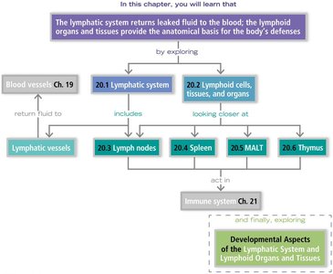
The lymphatic system is a vital component of the circulatory and immune systems, responsible for returning leaked fluid from blood vessels back to the blood and providing the anatomical basis for the body's defenses. It consists of lymphatic vessels, lymph, and lymphoid organs and tissues.
Lymphatic vessels: Network that transports lymph fluid.
Lymph: Fluid within lymphatic vessels, derived from interstitial fluid.
Lymph nodes: Structures that cleanse lymph and house immune cells.
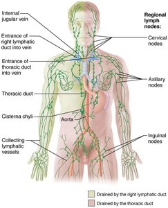
Anatomy of the Lymphatic System
The lymphatic system includes a network of vessels and several organs that play key roles in immunity and fluid balance.
Lymphatic vessels: Collect and return interstitial fluid to the bloodstream.
Major lymphoid organs: Spleen, thymus, tonsils, lymph nodes, and other tissues.
Regional lymph nodes: Cervical, axillary, and inguinal nodes filter lymph from specific body regions.
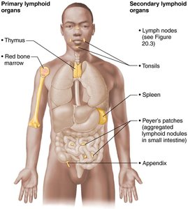
Primary and Secondary Lymphoid Organs
Lymphoid organs are classified as primary (where lymphocytes mature) and secondary (where immune responses are initiated).
Primary: Thymus and red bone marrow.
Secondary: Lymph nodes, tonsils, spleen, Peyer's patches, and appendix.
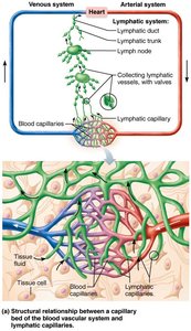
Structural Relationship: Blood and Lymphatic Capillaries
Lymphatic capillaries are closely associated with blood capillaries, facilitating the exchange and return of fluids.
Lymphatic capillaries collect excess tissue fluid and return it to the venous system.
Valves in lymphatic vessels ensure one-way flow toward the heart.
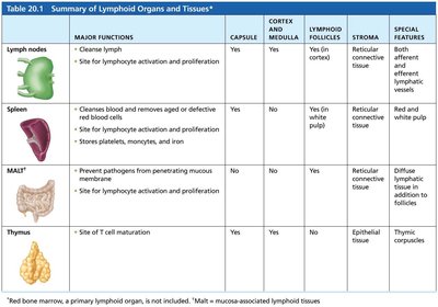
Summary Table: Lymphoid Organs and Tissues
Organ | Major Functions | Capsule | Cortex & Medulla | Lymphoid Follicles | Stroma | Special Features |
|---|---|---|---|---|---|---|
Lymph nodes | Cleanse lymph; site for lymphocyte activation and proliferation | Yes | Yes | On cortex | Reticular connective tissue | Both afferent and efferent lymphatic vessels |
Spleen | Cleanses blood, removes aged/defective RBCs; stores platelets, monocytes, iron | Yes | Yes | In white pulp | Reticular connective tissue | Red and white pulp |
MALT* | Prevent pathogens from penetrating mucosal surfaces; site for lymphocyte activation and proliferation | No | No | Yes | Diffuse lymphoid tissue | Scattered throughout mucosa |
Thymus | Site of T cell maturation | Yes | Yes | No | Epithelial tissue | Thymic corpuscles |
The Immune System
Overview and Organization
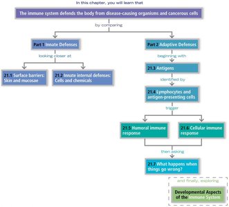
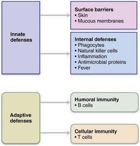
The immune system defends the body from disease-causing organisms and cancerous cells. It is divided into innate (nonspecific) and adaptive (specific) defenses.
Innate defenses: Surface barriers (skin, mucous membranes) and internal defenses (phagocytes, NK cells, inflammation, antimicrobial proteins, fever).
Adaptive defenses: Humoral immunity (B cells, antibodies) and cellular immunity (T cells).
Defense Mechanisms: Lines of Defense
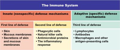
The immune system operates through three lines of defense:
First line: Skin, mucous membranes, and their secretions.
Second line: Phagocytic cells, NK cells, antimicrobial proteins, inflammatory response.
Third line: Lymphocytes, antibodies, antigen-presenting cells.
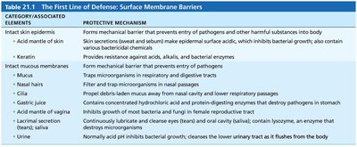
First Line of Defense: Surface Membrane Barriers
Surface barriers prevent entry of pathogens and harmful substances.
Skin: Mechanical barrier; acid mantle inhibits bacterial growth.
Mucous membranes: Trap microorganisms; cilia move debris out.
Secretions: Include enzymes, acids, and lysozyme that destroy pathogens.
Category/Element | Protective Mechanism |
|---|---|
Intact skin epidermis | Mechanical barrier; acid mantle inhibits bacteria |
Keratin | Resists acids, alkalis, bacterial enzymes |
Mucus | Traps microorganisms |
Gastric juice | Destroys pathogens in stomach |
Saliva | Contains lysozyme, cleanses oral cavity |
Urine | Acidic pH inhibits bacterial growth |
Second Line of Defense: Innate Cellular and Chemical Defenses
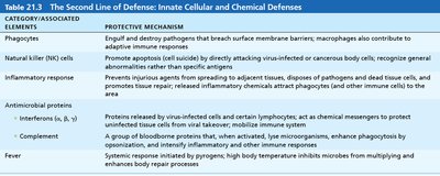
Internal defenses are activated when pathogens breach surface barriers.
Phagocytes: Engulf and destroy pathogens.
Natural killer (NK) cells: Induce apoptosis in infected or cancerous cells.
Inflammatory response: Localizes infection, disposes of debris, and initiates repair.
Antimicrobial proteins: Interferons and complement proteins enhance defense.
Fever: Inhibits microbial growth and accelerates repair.
Element | Protective Mechanism |
|---|---|
Phagocytes | Engulf and destroy pathogens; contribute to adaptive immunity |
NK cells | Induce apoptosis; recognize abnormal cells |
Inflammatory response | Localizes infection, disposes debris, attracts immune cells |
Interferons | Protect cells from viral infection |
Complement | Enhances phagocytosis, inflammation, cell lysis |
Fever | Inhibits microbes, enhances repair |
Phagocytosis: Mechanism and Events
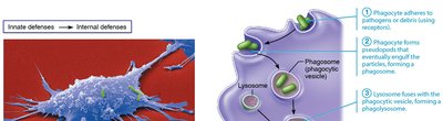
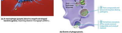
Phagocytes, such as macrophages and neutrophils, play a central role in innate immunity by engulfing pathogens.
Phagocyte adheres to pathogen using receptors.
Engulfment forms a phagosome.
Lysosome fuses with phagosome, forming a phagolysosome.
Enzymes and toxic compounds destroy the pathogen.
Residual material is exocytosed.
Inflammation: Acute and Chronic
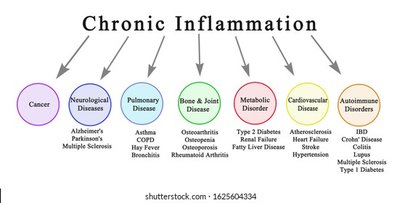
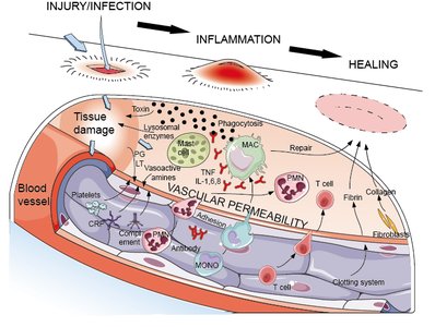
Inflammation is a localized response to tissue injury or infection, characterized by redness, heat, swelling, pain, and loss of function.
Prevents spread of damaging agents.
Disposes of cell debris and pathogens.
Alerts adaptive immune system.
Sets the stage for tissue repair.
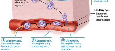
Phagocyte Mobilization During Inflammation
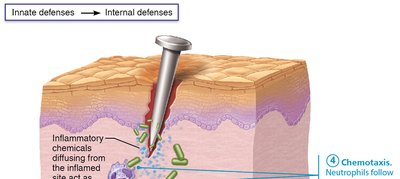
The mobilization of phagocytes involves several steps:
Leukocytosis: Increased production of neutrophils.
Margination: Neutrophils cling to capillary walls.
Diapedesis: Neutrophils squeeze through capillary walls.
Chemotaxis: Neutrophils follow chemical signals to the site of injury.
Complement System: Activation and Function
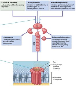
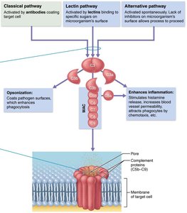
The complement system consists of ~20 blood proteins that circulate in inactive form and are activated via three pathways: classical, lectin, and alternative.
Classical pathway: Activated by antibodies binding to target cells.
Lectin pathway: Activated by lectins binding to sugars on microorganisms.
Alternative pathway: Activated spontaneously on microbial surfaces.
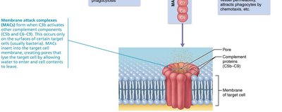
All pathways converge at C3, leading to opsonization, enhanced inflammation, and cell lysis via membrane attack complexes (MAC). 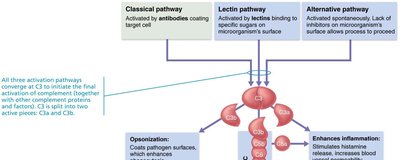
Fever: Systemic Response
Fever is an abnormally high body temperature induced by pyrogens released from leukocytes and macrophages.
Inhibits microbial growth by sequestering iron and zinc.
Increases metabolic rate, accelerating tissue repair.
Adaptive (Specific) Immunity
Overview and Characteristics
Adaptive immunity provides specific, systemic, and memory-based protection against pathogens. It consists of two overlapping arms: humoral (antibody-mediated) and cellular (cell-mediated) immunity.
Humoral immunity: B cells produce antibodies that target extracellular pathogens.
Cellular immunity: T cells target infected or abnormal cells.
Humoral Immunity: B Cells and Antibodies
Humoral immunity involves the production of antibodies by B cells.
B cells mature in bone marrow and migrate to lymphoid organs.
Upon encountering an antigen, B cells undergo clonal selection and differentiate into plasma cells, which secrete antibodies.
Antibodies neutralize, agglutinate, precipitate antigens, and activate complement.
Antibody Structure and Classes
Antibodies are Y-shaped molecules composed of two heavy and two light chains, with variable regions forming antigen-binding sites.
Constant regions determine antibody class (IgM, IgA, IgD, IgG, IgE).
Classes differ in function and location.
Lymphocyte Development, Maturation, and Activation
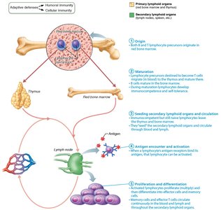
Lymphocytes originate in red bone marrow, mature in primary lymphoid organs, and are seeded in secondary lymphoid organs. Activation occurs upon antigen encounter, followed by proliferation and differentiation.
Origin: Precursors in bone marrow.
Maturation: T cells in thymus, B cells in bone marrow.
Seeding: Secondary lymphoid organs.
Activation: Antigen encounter.
Proliferation: Clonal expansion and differentiation.
Cellular Immunity: T Cells
Cellular immunity is mediated by T lymphocytes, which recognize antigens presented by MHC proteins.
Helper T cells (TH): Stimulate B cells and other T cells.
Cytotoxic T cells (TC): Kill infected or cancerous cells directly.
Memory T cells: Provide long-term immunity.
Regulatory T cells: Modulate immune response.
Mechanisms of Target Cell Killing by T Cells
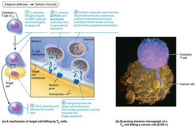
Cytotoxic T cells kill target cells by releasing perforin and granzymes, which induce apoptosis.
Perforin forms pores in target cell membrane.
Granzymes enter and trigger apoptosis.
Summary: Humoral Immunity Pathways
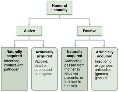
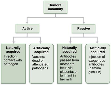
Humoral immunity can be acquired actively (infection or vaccination) or passively (maternal antibodies or injection of antibodies).
Active: Naturally (infection) or artificially (vaccination).
Passive: Naturally (maternal antibodies) or artificially (injection).
Antigen-Antibody Complex: Effects
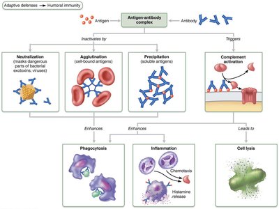
Antigen-antibody complexes inactivate antigens and trigger complement activation, enhancing phagocytosis, inflammation, and leading to cell lysis.
Neutralization: Masks dangerous parts of antigens.
Agglutination: Clumps cell-bound antigens.
Precipitation: Aggregates soluble antigens.
Complement activation: Leads to cell lysis.
Key Terms and Concepts
Lymph: Fluid in lymphatic vessels.
Antigen: Substance that triggers an immune response.
Antibody: Protein produced by B cells that binds antigens.
MHC: Major histocompatibility complex, presents antigens to T cells.
Phagocytosis: Cellular process of engulfing pathogens.
Complement: System of proteins enhancing immunity.
Inflammation: Localized response to injury or infection.
Fever: Systemic response to infection.
Summary
The lymphatic and immune systems work together to maintain fluid balance and defend the body against pathogens. The lymphatic system returns leaked fluid to the blood and provides sites for immune cell activation. The immune system employs both innate and adaptive mechanisms to protect against infection and disease, utilizing a variety of cells, proteins, and processes to ensure effective defense and repair.
