 Indietro
Lymphatic System and Innate Immunity: Structure and Function
Guida di studio - Note intelligenti
 Appunti personalizzati basati sui tuoi materiali, ampliati con definizioni chiave, esempi e contesto.
Lymphatic System: Structure and Function
Overview of the Lymphatic System
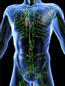
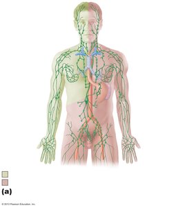
The lymphatic system is a vital component of the circulatory and immune systems, responsible for returning interstitial fluid to the bloodstream and providing a structural basis for immune defense. It consists of a network of lymphatic vessels, lymph (the fluid within these vessels), and lymphoid organs and tissues.
Lymphatic vessels collect and transport lymph toward the heart in a one-way system.
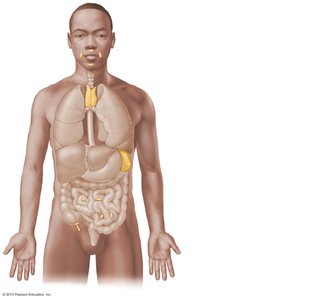
Lymphoid organs and tissues include the spleen, thymus, tonsils, lymph nodes, and other tissues that house immune cells.
These structures provide sites for immune cell proliferation and surveillance.
Functions of the Lymphatic System
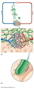
Returns interstitial fluid and leaked plasma proteins to the blood (about 3L/day).
Transports dietary fats from the intestine to the blood via specialized lymphatic capillaries called lacteals.
Provides a route for immune cells to monitor and respond to pathogens.
Lymphatic Vessels and Circulation
Lymphatic vessels are organized into a hierarchy:
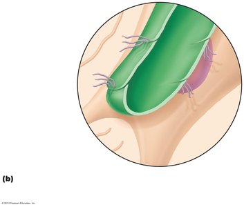
Lymphatic capillaries: Highly permeable, blind-ended tubes that absorb proteins, cell debris, and pathogens. Their endothelial cells overlap to form one-way minivalves, and they are anchored by collagen filaments.
Collecting lymphatic vessels: Similar to veins but with thinner walls and more valves.
Lymphatic trunks: Formed by the union of collecting vessels; major trunks include lumbar, bronchomediastinal, subclavian, jugular, and intestinal trunks.
Lymphatic ducts: The right lymphatic duct drains the right upper arm and right side of the head and thorax; the thoracic duct drains the rest of the body. Both empty into the venous circulation at the junction of the internal jugular and subclavian veins.
Lymph Transport Mechanisms
Propelled by skeletal muscle contraction, thoracic pressure changes during breathing, pulsations of nearby arteries, and smooth muscle contractions in vessel walls.
Valves prevent backflow, ensuring unidirectional flow toward the heart.
Lymphoid Cells and Tissues
Lymphoid cells are essential for immune defense and include:
Lymphocytes: Main immune cells, divided into T cells (mediate cellular immunity) and B cells (produce antibodies for humoral immunity).
Macrophages: Phagocytize foreign substances and help activate T cells.
Dendritic cells: Capture antigens and deliver them to lymph nodes, activating T cells.
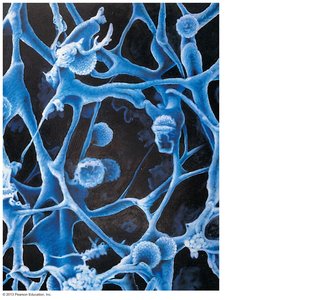
Reticular cells: Produce the reticular fiber stroma that supports other cells in lymphoid organs.
Lymphoid Tissue Types
Diffuse lymphoid tissue: Loosely arranged lymphoid cells and reticular fibers found in most organs.
Lymphoid follicles (nodules): Solid, spherical bodies of tightly packed lymphoid cells; contain germinal centers with proliferating B cells. Found in Peyer's patches and the appendix.
Lymphoid Organs
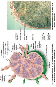
Lymph nodes: Principal lymphoid organs that filter lymph and activate the immune system. Located in clusters along lymphatic vessels, especially in the inguinal, axillary, and cervical regions.
Spleen: Largest lymphoid organ; site of lymphocyte proliferation, immune surveillance, and blood cleansing. Stores breakdown products of RBCs, platelets, and monocytes.
Thymus: Site of T cell maturation; most active during childhood and atrophies with age. Differs from other lymphoid organs by lacking B cells and not directly fighting antigens.
MALT (Mucosa-associated lymphoid tissue): Lymphoid tissues in mucous membranes throughout the body, including tonsils, Peyer's patches, and the appendix. Protects against pathogens entering through mucosa.
Innate Immunity: Nonspecific Defenses
Overview of Immunity
Immunity is the body's ability to resist disease. The immune system is divided into two main branches:
Innate (nonspecific) immunity: Provides immediate, general defense against pathogens.
Adaptive (specific) immunity: Targets specific pathogens and provides long-lasting protection.
Innate Defenses: Surface Barriers
Skin and mucous membranes act as physical barriers to most microorganisms.
Protective chemicals include the acid mantle, lysozyme, defensins, and other antimicrobial substances.
Respiratory system modifications (mucus, cilia) help trap and remove pathogens.
Innate Defenses: Internal Defenses
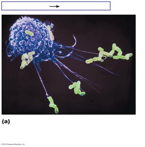
Phagocytes: Neutrophils and macrophages ingest and destroy pathogens.
Natural killer (NK) cells: Attack cells lacking "self" markers, inducing apoptosis in cancer and virus-infected cells.
Antimicrobial proteins: Interferons and complement proteins inhibit microbial growth and enhance immune responses.
Fever: Elevated body temperature inhibits pathogen growth and speeds up tissue repair.
Inflammation: Local response to injury that prevents spread of damage, disposes of pathogens, and sets the stage for repair.
Phagocytosis Mechanism
Phagocytes adhere to pathogens, often enhanced by opsonization (coating by antibodies or complement proteins).
Pathogen is engulfed into a phagosome, which fuses with a lysosome to form a phagolysosome.
Lysosomal enzymes digest the pathogen; indigestible material is exocytosed.
Inflammatory Response
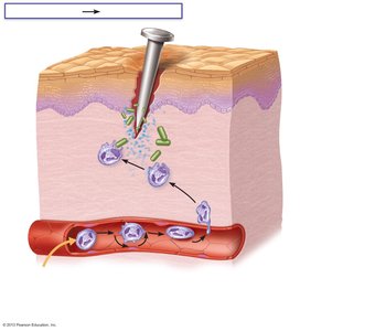
Triggered by tissue injury; characterized by redness, heat, swelling, pain, and sometimes loss of function.
Chemical mediators (histamine, kinins, prostaglandins, complement) cause vasodilation, increased permeability, and attract leukocytes.
Leukocyte mobilization involves leukocytosis, margination, diapedesis, and chemotaxis.
Antimicrobial Proteins: Interferons and Complement System
Interferons (IFNs): Proteins released by virus-infected cells to warn neighboring cells, inducing production of antiviral proteins and activating NK cells.
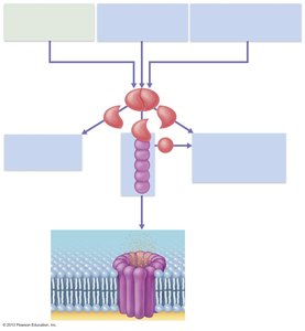
Complement system: A group of about 20 plasma proteins that enhance inflammation, promote phagocytosis (opsonization), and cause cell lysis via the membrane attack complex (MAC).
Complement activation occurs via three pathways: classical (antibody-mediated), lectin, and alternative (spontaneous).
Summary Table: Major Lymphatic Structures and Functions
Structure | Main Function |
|---|---|
Lymphatic vessels | Return interstitial fluid to blood; transport lymph |
Lymph nodes | Filter lymph; activate immune response |
Spleen | Immune surveillance; cleanses blood; stores blood components |
Thymus | T cell maturation |
MALT | Protects mucosal surfaces from pathogens |
Additional info: The lymphatic and innate immune systems are closely integrated with the adaptive immune system, which provides specific, long-term immunity through the action of T and B lymphocytes. Understanding the structure and function of lymphatic organs is foundational for comprehending immune responses and disease resistance.
