 Indietro
Major Skeletal Muscles of the Human Body: Structure, Function, and Location
Guida di studio - Note intelligenti
 Appunti personalizzati basati sui tuoi materiali, ampliati con definizioni chiave, esempi e contesto.
Major Skeletal Muscles of the Body
Overview of Skeletal Muscles
The human body contains over 600 skeletal muscles, which are grouped by their function and anatomical location. Understanding the origin, insertion, and action of each muscle is essential for comprehending movement and musculoskeletal anatomy.
Origin: The fixed attachment point of the muscle, usually proximal.
Insertion: The movable attachment point, usually distal.
Action: The movement produced when the muscle contracts.
Muscle Naming: Names often indicate location, function, shape, or attachments.
Tips for Learning Muscles:
Analyze muscle names for clues about their function or location.
Use anatomical figures to identify muscles visually.
Relate muscle attachments to their actions.
Practice movements to understand muscle function.
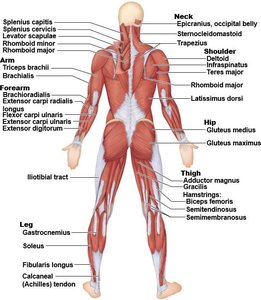
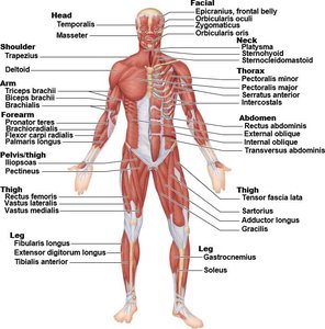
Muscles of the Head and Neck
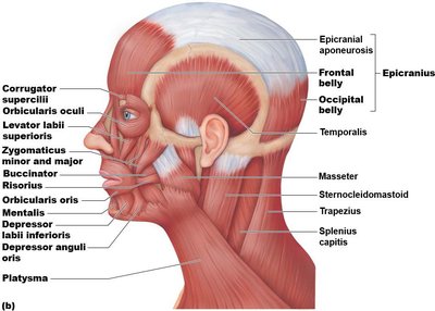
Facial Muscles
Facial muscles are responsible for expressions, mastication, and movements of the head and neck. They are often attached to skin, allowing for subtle movements.
Occipitofrontalis: Consists of frontal and occipital bellies connected by an aponeurosis; moves scalp, raises eyebrows, wrinkles forehead.
Orbicularis oculi: Sphincter muscle surrounding the eye; closes eyelids, enables blinking and squinting.
Zygomaticus: Extends from cheekbone to mouth corner; raises lateral corners of mouth (smiling).
Orbicularis oris: Encircles the mouth; puckers lips.
Buccinator: Compresses cheek, assists in whistling, sucking, and holds food between teeth during chewing.
Platysma: Tenses skin of neck; superficial sheet-like muscle.
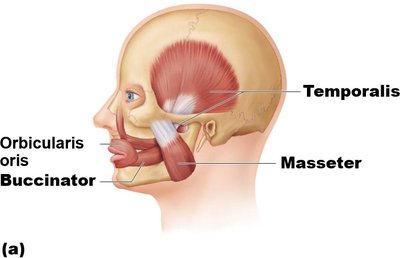
Muscles of Mastication
Masseter: Elevates mandible; primary muscle for closing jaw during chewing.
Temporalis: Elevates mandible; assists in closing jaw.
Neck Muscles
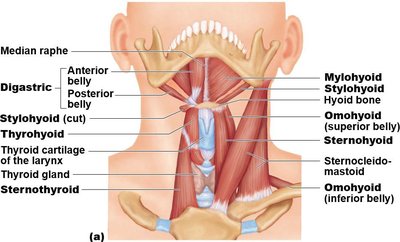
Sternocleidomastoid: Flexes and rotates neck; each side rotates head toward opposite shoulder and tilts head to its own side.
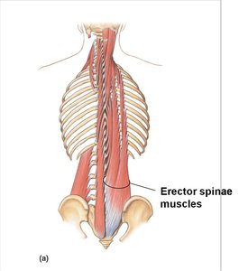
Erector spinae: Prime movers of back extension; includes iliocostalis, longissimus, and spinalis muscles. Provides resistance for bending forward and returns body to erect position.
Muscles of the Thorax: Breathing
Intercostal Muscles
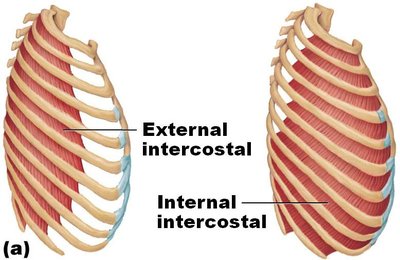
Intercostal muscles are located between the ribs and play a crucial role in respiration.
External intercostals: Elevate rib cage, aiding inspiration.
Internal intercostals: Depress rib cage, aiding forced expiration.
Diaphragm
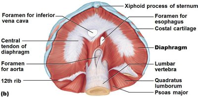
The diaphragm is a dome-shaped muscle forming the floor of the thoracic cavity. It is the primary muscle of inspiration.
Origin: Xiphoid process, costal cartilages, lumbar vertebrae.
Insertion: Central tendon of diaphragm.
Action: Flattens during contraction, increasing thoracic volume and drawing air into lungs.
Muscles of the Abdominal Wall
Abdominal Muscles
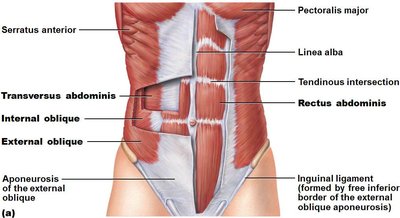
Abdominal muscles support trunk movement, protect internal organs, and assist in respiration.
Rectus abdominis: Flexes and rotates lumbar region; segmented by tendinous intersections.
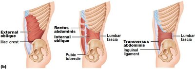
External oblique: Largest and most superficial lateral muscle; flexes vertebral column, rotates trunk, and produces lateral flexion.
Internal oblique: Deep to external oblique; similar actions.
Transversus abdominis: Deepest muscle; compresses abdominal contents.
Superficial Muscles of the Anterior and Posterior Thorax
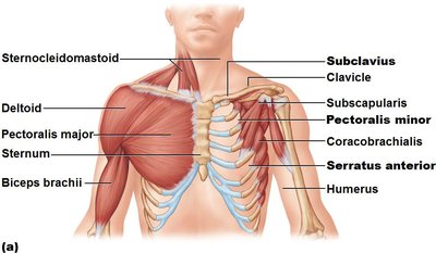
Anterior Thorax Muscles
Pectoralis major: Flexes shoulder, medially rotates and adducts arm.
Pectoralis minor: Draws scapula forward and downward.
Serratus anterior: Protracts scapula, holds it against chest wall.
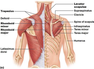
Posterior Thorax Muscles
Trapezius: Elevates, retracts, and rotates scapula.
Levator scapulae: Elevates scapula.
Rhomboids (major and minor): Retract scapula; located deep to trapezius and inferior to levator scapulae.
Summary Table: Major Muscles, Origins, Insertions, and Actions
Muscle Name | Origin | Insertion | Action |
|---|---|---|---|
Occipitofrontalis | Occipital/frontal bones | Aponeurosis | Moves scalp, raises eyebrows |
Orbicularis oculi | Frontal/maxillary bones | Eyelid tissue | Closes eye |
Zygomaticus | Zygomatic bone | Corner of mouth | Smiling |
Orbicularis oris | Maxilla/mandible | Angles of mouth | Puckers lips |
Buccinator | Molar region of maxilla/mandible | Orbicularis oris | Compresses cheek |
Platysma | Fascia of chest | Lower margin of mandible | Tenses neck skin |
Masseter | Zygomatic arch/bone | Mandible | Elevates mandible |
Temporalis | Temporal bone | Coronoid process of mandible | Elevates mandible |
Sternocleidomastoid | Sternum/clavicle | Mastoid process | Flexes/rotates neck |
Erector spinae | Vertebrae/ribs | Vertebrae/ribs | Extends back |
External intercostals | Rib above | Rib below | Elevates rib cage |
Internal intercostals | Rib below | Rib above | Depresses rib cage |
Diaphragm | Xiphoid process, ribs, vertebrae | Central tendon | Inspiration |
Rectus abdominis | Pubic bone | Xiphoid process | Flexes/rotates lumbar region |
External oblique | Lower ribs | Linea alba, iliac crest | Flexes/rotates trunk |
Internal oblique | Iliac crest | Linea alba, lower ribs | Flexes/rotates trunk |
Transversus abdominis | Iliac crest, lower ribs | Linea alba | Compresses abdomen |
Pectoralis major | Sternum, clavicle | Humerus | Flexes, adducts, rotates arm |
Pectoralis minor | Ribs 3-5 | Coracoid process | Draws scapula forward/down |
Serratus anterior | Ribs 1-8 | Scapula | Protracts scapula |
Trapezius | Occipital bone, C7, thoracic vertebrae | Scapula, clavicle | Elevates/retracts scapula |
Levator scapulae | C1-C4 | Scapula | Elevates scapula |
Rhomboids | C7-T5 | Scapula | Retracts scapula |
Key Concepts and Applications
Muscle Attachments: The origin and insertion determine the muscle's action.
Muscle Actions: Muscles work in groups to produce complex movements.
Clinical Relevance: Understanding muscle anatomy is essential for diagnosing injuries and planning rehabilitation.
Example: Flexion of the Neck
Simultaneous contraction of both sternocleidomastoid muscles flexes the neck, while contraction of one side rotates the head toward the opposite shoulder.
Example: Breathing
The diaphragm contracts and flattens during inspiration, increasing thoracic volume. External intercostals elevate the rib cage, while internal intercostals depress it during forced expiration.
Example: Trunk Rotation
External and internal oblique muscles contract individually to rotate the trunk and produce lateral flexion.
Additional info: Expanded explanations and clinical context were added to ensure completeness and academic quality.
