 Indietro
Major Veins of the Systemic Circulation
Guida di studio - Note intelligenti
 Appunti personalizzati basati sui tuoi materiali, ampliati con definizioni chiave, esempi e contesto.
Major Veins of the Systemic Circulation
The systemic venous system returns deoxygenated blood from the body tissues back to the right atrium of the heart. Understanding the major veins and their regional organization is essential for students of anatomy and physiology, as these vessels are critical for clinical procedures and understanding circulatory physiology.
Venous Drainage of the Head, Neck, and Brain
The veins of the head and neck collect blood from the brain, scalp, and face, channeling it toward the heart. The internal and external jugular veins are the primary conduits for this region.
Internal Jugular Vein: Drains blood from the brain, face, and neck; joins the subclavian vein to form the brachiocephalic vein.
External Jugular Vein: Drains superficial regions of the scalp and face.
Vertebral Vein: Drains the cervical spinal cord and posterior skull.
Other Key Veins: Ophthalmic, facial, occipital, posterior auricular, and superficial temporal veins contribute to the venous return from the head.
Venous Sinuses: The brain is drained by dural venous sinuses, which empty into the internal jugular vein.
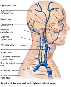
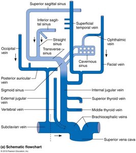
Example: The internal jugular vein is commonly used for central venous catheterization in clinical practice.
Veins of the Thorax and Upper Limb
The veins of the thorax and upper limb return blood from the chest wall, upper extremities, and portions of the back to the heart. The subclavian and brachiocephalic veins are major vessels in this region.
Subclavian Vein: Continuation of the axillary vein; receives blood from the upper limb.
Brachiocephalic Vein: Formed by the union of the subclavian and internal jugular veins; left and right brachiocephalic veins merge to form the superior vena cava.
Major Upper Limb Veins: Cephalic, basilic, median cubital, and brachial veins drain the arm and forearm.
Thoracic Veins: Azygos system drains the thoracic wall and provides collateral circulation between the superior and inferior vena cava.
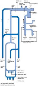
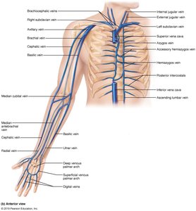
Example: The median cubital vein is often used for venipuncture (drawing blood).
Veins of the Abdomen
Abdominal veins drain blood from the abdominal organs and walls. The inferior vena cava is the main vessel returning blood from the lower body to the heart.
Inferior Vena Cava: Formed by the union of the common iliac veins; receives blood from the lower limbs, pelvis, and abdominal wall.
Renal Veins: Drain the kidneys directly into the inferior vena cava.
Hepatic Veins: Drain blood from the liver into the inferior vena cava.
Hepatic Portal System: Blood from the digestive organs is first filtered through the liver via the hepatic portal vein before entering the systemic circulation.
Other Key Veins: Suprarenal, gonadal, lumbar, and phrenic veins contribute to abdominal venous return.
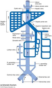
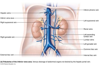
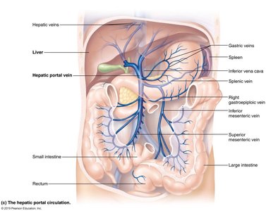
Example: The hepatic portal vein carries nutrient-rich blood from the gastrointestinal tract to the liver for processing.
Veins of the Lower Limb
The veins of the lower limb return blood from the foot, leg, and thigh to the heart. These veins are divided into superficial and deep systems, which communicate via perforating veins.
Superficial Veins: Great saphenous and small saphenous veins are the major superficial veins; often used for grafts in coronary bypass surgery.
Deep Veins: Include the anterior tibial, posterior tibial, fibular (peroneal), popliteal, and femoral veins.
Venous Flow: Blood from the foot drains into the deep and superficial veins, which ascend and eventually form the femoral vein, continuing as the external iliac vein.
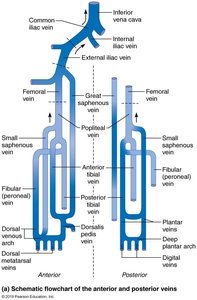
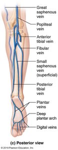
Example: The great saphenous vein is the longest vein in the body and is frequently harvested for use in coronary artery bypass grafting.
Summary Table: Major Systemic Veins and Their Regions
Region | Major Veins | Key Tributaries |
|---|---|---|
Head & Neck | Internal jugular, external jugular, vertebral | Facial, occipital, superficial temporal |
Thorax & Upper Limb | Subclavian, brachiocephalic, azygos | Cephalic, basilic, median cubital, brachial |
Abdomen | Inferior vena cava, hepatic portal | Renal, hepatic, suprarenal, gonadal |
Lower Limb | Femoral, great saphenous, small saphenous | Anterior tibial, posterior tibial, fibular |
Additional info: The systemic veins are often paired with arteries of the same name, but their courses and tributaries can differ significantly. Understanding venous anatomy is crucial for clinical procedures such as venipuncture, central line placement, and the management of venous diseases.
