 Indietro
Membrane Transport & Excitable Cells: Neuron Physiology and Synaptic Transmission
Guida di studio - Note intelligenti
 Appunti personalizzati basati sui tuoi materiali, ampliati con definizioni chiave, esempi e contesto.
Membrane Transport & Excitable Cells
Overview of Neurons and Neuroglia
Neurons are highly specialized cells responsible for transmitting electrical signals throughout the nervous system. They require support from neuroglial cells, which maintain the environment and provide structural and metabolic support. Neuroglia are classified based on their location in the central nervous system (CNS) or peripheral nervous system (PNS).
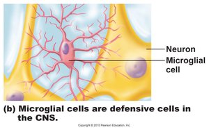
Microglia: Protective cells in the CNS that monitor neuron health and can transform into macrophages to engulf microorganisms and debris.
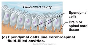
Ependymal cells: Line the cavities of the brain and spinal cord, forming a barrier between cerebrospinal fluid (CSF) and CNS tissue; their cilia help circulate CSF.
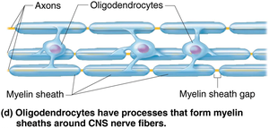
Oligodendrocytes: Provide myelin sheaths to CNS neurons, facilitating rapid signal transmission.
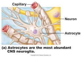
Astrocytes: Star-shaped, most abundant CNS neuroglia; anchor neurons to capillaries, facilitate nutrient exchange, guide neuron migration, and clean up excess neurotransmitters and ions.
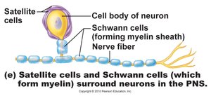
Satellite cells: Surround neuron cell bodies in PNS ganglia, performing functions similar to astrocytes.
Schwann cells: Form myelin sheaths around larger PNS neurons and are vital for peripheral nerve regeneration.
Structural and Functional Classification of Neurons
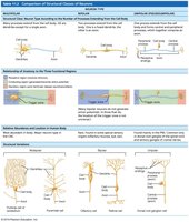
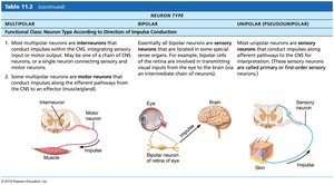
Neurons are classified based on the number of processes extending from the cell body (structural) and the direction of impulse conduction (functional).
Structural: Multipolar, bipolar, and unipolar neurons.
Functional: Sensory (afferent), motor (efferent), and association (interneurons).
Resting Membrane Potential (RMP)
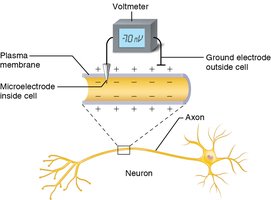
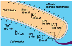
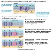
The resting membrane potential is the voltage difference across the plasma membrane when the cell is at rest, typically around -70 mV. This negativity is due to the differential permeability of the membrane to sodium (Na+) and potassium (K+) ions, and the activity of the Na+/K+ pump.
Key cations: K+ is primarily inside cells; Na+ is primarily outside.
Leaky channels: The membrane is more permeable to K+ than Na+, resulting in a net loss of positive charge from the cell.
Na+/K+ pump: Moves 3 Na+ out for every 2 K+ in, maintaining the gradient.
Ion Channels in Neuronal Membranes
Channels in the plasma membrane allow ions to move passively in response to electrochemical gradients. They are classified as:
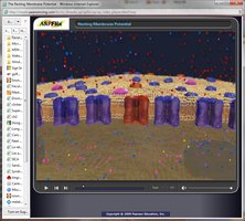
Leakage channels: Always open, responsible for resting membrane potential.
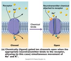
Gated channels: Require a signal to open/close; can be chemically-gated (neurotransmitter/hormone) or voltage-gated (change in membrane potential).
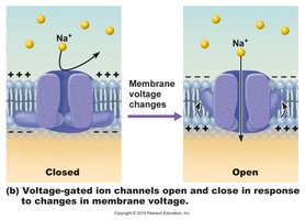
Changes in Membrane Potential: Depolarization and Hyperpolarization
Neurons and muscle cells communicate by changing membrane potentials, producing two types of signals: graded potentials and action potentials.
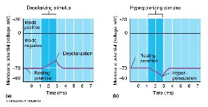
Depolarization: Increases the probability of producing nerve impulses; membrane potential becomes less negative.
Hyperpolarization: Decreases the probability; membrane potential becomes more negative.
Graded Potentials
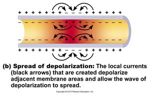
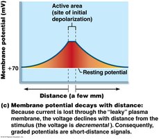
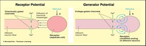
Graded potentials are short-lived changes in membrane potential, produced in dendrites. Their magnitude depends on the strength of the stimulus, and the current decreases with distance traveled (decremental).
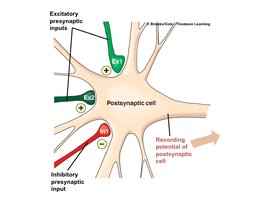
Types: Generator/receptor potentials (triggered by external stimuli) and postsynaptic potentials (triggered by neurotransmitter release).
Action Potentials (AP)
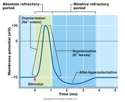
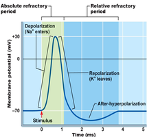
An action potential is a brief reversal of membrane potential (from -70 mV to +30 mV), generated at the axon hillock and propagated along the axon. APs do not decrease in amplitude with distance and are generated by voltage-gated channels.
Depolarizing phase: Transient increase in Na+ permeability; voltage-gated Na+ channels open.
Threshold: The minimum membrane potential required to trigger an AP (typically -55 to -50 mV).
Repolarizing phase: Decrease in Na+ permeability and increase in K+ permeability; K+ leaves the cell, restoring negativity.
After-hyperpolarization: Due to slow closure of K+ channels.
Na+/K+ pump: Restores ion gradients after AP.
Absolute and Relative Refractory Periods
During the absolute refractory period, Na+ channels are open and a second depolarization is impossible. During the relative refractory period, Na+ channels have closed but K+ channels are open; a very strong stimulus can trigger another AP.
Propagation of Action Potentials
Action potentials propagate along the axon, traversing its length to signal the next neuron. Propagation is unidirectional due to refractory periods, ensuring that Na+ channels cannot reopen immediately.
Threshold and All-or-None Phenomenon
At threshold, outward K+ current equals inward Na+ current.
APs are all-or-none events: once threshold is reached, the AP is always the same size.
Stimulus intensity is coded by AP frequency, not amplitude.
Myelin Sheaths and Saltatory Conduction
Myelin sheaths, produced by Schwann cells (PNS) and oligodendrocytes (CNS), act as insulators and increase the rate of impulse propagation (up to 150x).
Nodes of Ranvier (gaps in myelin) allow saltatory conduction, where APs jump from node to node.
The Synapse and Synaptic Transmission
A synapse is the junction between two neurons or a neuron and an effector. Most neurons are both presynaptic and postsynaptic. There are two types of synapses: electrical and chemical.
Electrical synapses: Direct current flow via gap junctions; rapid transmission and synchronization.
Chemical synapses: Neurotransmitters released from axonal terminals bind to receptors on the postsynaptic membrane, opening ion channels.
Mechanism of Synaptic Communication
Ca2+ gates open in presynaptic terminal.
Neurotransmitter release.
Neurotransmitter binds to postsynaptic receptors.
Ion channels open in postsynaptic membrane.
Termination of Synaptic Transmission
Degradation by enzymes (e.g., acetylcholine).
Reuptake by presynaptic terminal (e.g., norepinephrine).
Diffusion away from synaptic site (e.g., nitric oxide).
Postsynaptic Potentials (PSPs)
Excitatory postsynaptic potentials (EPSPs): NT binding causes membrane depolarization; if strong enough, can trigger an AP.
Inhibitory postsynaptic potentials (IPSPs): NT binding causes membrane hyperpolarization, making AP generation less likely.
Summation of PSPs
Temporal summation: Multiple signals in quick succession.
Spatial summation: Multiple signals from different locations.
Most effective synapses are closest to the axon hillock.
Neurotransmitter Classification
Chemical: Acetylcholine, biogenic amines (dopamine, norepinephrine, epinephrine, serotonin, histamine), amino acids (glutamate, aspartate, glycine, GABA), neuropeptides (substance P, endorphins), purines (ATP, adenosine), gases & lipids (NO, CO, H2S), endocannabinoids.
Functional: Excitatory vs. inhibitory (depolarization vs. hyperpolarization); direct vs. indirect (channel opening vs. second messenger activation).
Neural Circuits
Neural processing can be serial (e.g., reflexes) or parallel (e.g., complex behaviors). Neurons in the CNS are organized into pools to handle information processing complexity.
Neuroglia Type | Location | Function |
|---|---|---|
Astrocytes | CNS | Support, nutrient exchange, clean up environment |
Microglia | CNS | Defense, phagocytosis |
Ependymal cells | CNS | Line cavities, circulate CSF |
Oligodendrocytes | CNS | Myelinate axons |
Satellite cells | PNS | Support neuron cell bodies |
Schwann cells | PNS | Myelinate axons, regeneration |
Neuron Type | Structure | Function |
|---|---|---|
Multipolar | Many processes | Motor, interneurons |
Bipolar | Two processes | Sensory (special senses) |
Unipolar | One process | Sensory (general) |
Additional info: The notes expand on brief points to provide full academic context, including definitions, examples, and tables for classification. All images included are directly relevant to the adjacent content, reinforcing key concepts in neuron physiology and synaptic transmission.
