 Indietro
Membrane Transport & Excitable Cells: Neurons, Glia, and the Electrical Language of the Nervous System
Guida di studio - Note intelligenti
 Appunti personalizzati basati sui tuoi materiali, ampliati con definizioni chiave, esempi e contesto.
Membrane Transport & Excitable Cells
Introduction
This unit explores the structure and function of neurons and glial cells, the mechanisms of membrane transport, and the electrical properties that enable nerve impulse transmission. Understanding these concepts is fundamental to grasping how the nervous system communicates and controls body functions.
Anatomy of a Neuron
Regions and Functions of a Neuron
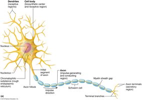
Neurons are specialized cells designed for rapid communication. Each region of a neuron has a distinct role in the reception, integration, propagation, and transmission of nerve impulses.
Reception: Dendrites and the cell body receive incoming signals, primarily neurotransmitters, from other neurons.
Integration: The axon hillock (initial segment) acts as the trigger zone, deciding whether to initiate an action potential.
Propagation: The axon conducts the action potential away from the soma toward the axon terminals.
Transmission: Axon terminals convert the electrical signal into a chemical one by releasing neurotransmitters.
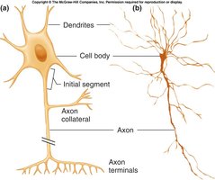
Cell Body (Soma/Perikaryon)
The cell body is the biosynthetic center of the neuron, containing the nucleus, nucleolus, Nissl bodies (rough ER), and neurofibrils (cytoskeleton). It is also a receptive region for synaptic input.
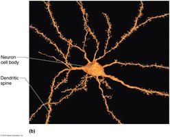
Dendrites
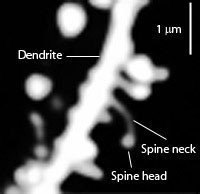
Dendrites are short, highly branched processes that serve as the main input region. They carry graded potentials toward the soma and are covered with dendritic spines, which are sites of most excitatory synapses and are involved in learning and memory.
Axon
The axon is a single, long process that propagates action potentials. It originates at the axon hillock and may branch into collaterals and terminal branches, ending in axon terminals that secrete neurotransmitters. Axonal transport moves materials between the soma and axon terminals.
Signature Properties of Neurons
Excitability: Ability to rapidly change membrane potential via gated channels.
Extreme Longevity: Neurons can function for a lifetime.
Amitotic: Most mature neurons do not divide.
High Metabolic Rate: Require constant oxygen and glucose.
Structural and Functional Classification
Multipolar: Many dendrites, one axon; most common (motor neurons, interneurons).
Bipolar: One dendrite, one axon; rare (retina, olfactory mucosa).
Unipolar: Single process splits into peripheral and central branches; sensory neurons.
Functionally, neurons are classified as sensory (afferent), interneurons, or motor (efferent) based on the direction of information flow.
Neuroglia (Glial Cells)
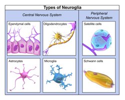
Types and Functions of Glial Cells
Glial cells support, insulate, and protect neurons. They are essential for nervous system function and outnumber neurons in the CNS.
Central Nervous System (CNS) | Peripheral Nervous System (PNS) |
|---|---|
Astrocytes | Satellite cells |
Microglia | Schwann cells |
Ependymal cells | |
Oligodendrocytes |
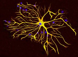
Astrocytes
Most abundant CNS glia; support neurons and link them to capillaries.
Regulate extracellular K+, recycle neurotransmitters, and help form the blood-brain barrier.
Modulate synaptic activity and form glial scars after injury.
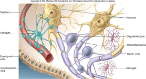
Microglia and Ependymal Cells
Microglia: Act as resident immune cells, phagocytosing debris and pathogens.
Ependymal cells: Line brain ventricles and central canal, circulate cerebrospinal fluid with cilia.

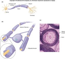
Oligodendrocytes vs. Schwann Cells
Oligodendrocytes (CNS): Form myelin sheaths around multiple axons.
Schwann cells (PNS): Myelinate single axons; aid in regeneration.
Myelin: Lipid-rich, insulating layer that increases conduction speed; nodes of Ranvier are gaps between myelin segments.
Resting Membrane Potential (RMP)
Electrical Properties of the Membrane
All cells maintain a voltage difference across their membrane, known as the resting membrane potential (RMP). In neurons, RMP is typically –70 mV.
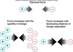
Voltage (Potential): Energy from charge separation (measured in mV).
Current: Flow of ions across the membrane.
Resistance: Hindrance to ion flow; lipid bilayer is an insulator, channels are conductors.
Ohm’s Law:
Ion Gradients and the Na+/K+ ATPase
Na+ and Cl– are higher outside; K+ is higher inside.
The Na+/K+ ATPase pumps 3 Na+ out and 2 K+ in per ATP, maintaining gradients.
Equilibrium Potentials and the Nernst Equation
Each ion has an equilibrium potential (Eion) where its electrical and chemical gradients balance. The Nernst equation calculates this:
EK ≈ –90 mV; ENa ≈ +60 mV
At rest, the membrane is more permeable to K+, so RMP is closer to EK.
Leak Channels and RMP
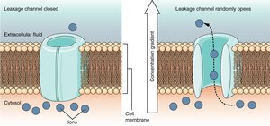
Leak channels are always open, allowing passive ion movement.
Resting membrane is ~25× more permeable to K+ than Na+.
RMP is set mainly by K+ gradient, with a small Na+ influence.
Changing the Signal: Graded and Action Potentials
Types of Ion Channels
Leak (non-gated): Always open; set resting potential.
Chemically (ligand-) gated: Open when neurotransmitter binds; found on dendrites and soma.
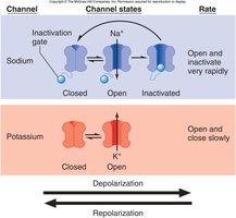
Voltage-gated: Open/close in response to membrane voltage; found on axons, generate action potentials.
Mechanically gated: Open in response to membrane deformation; found in sensory receptors.

Depolarization and Hyperpolarization
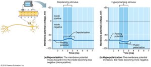
Depolarization: Membrane potential becomes less negative (e.g., Na+ influx).
Hyperpolarization: Membrane potential becomes more negative (e.g., K+ efflux or Cl– influx).
Graded Potentials
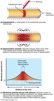
Local, variable changes in membrane potential.
Triggered by stimuli that open gated channels; size depends on stimulus strength.
Decay with distance due to current leakage.
Action Potentials
Action potentials are all-or-none, long-distance signals generated by voltage-gated channels in axons.
Resting: All voltage-gated channels closed; RMP maintained by leak channels.
Depolarization: Na+ channels open rapidly; Na+ influx causes upstroke.
Repolarization: Na+ channels inactivate; K+ channels open slowly; K+ efflux restores negativity.
Hyperpolarization: K+ channels close slowly, causing undershoot.
Refractory Periods
Absolute refractory period: No new action potential can be generated (Na+ channels inactivated).
Relative refractory period: Only a stronger stimulus can trigger another action potential (K+ channels still open).

Action Potential Propagation
Action potentials propagate as a wave of depolarization, regenerating at each patch of membrane. The refractory period ensures one-way travel.
Conduction velocity increases with axon diameter and myelination.
Saltatory conduction: In myelinated axons, action potentials jump between nodes of Ranvier, greatly increasing speed.
Clinical Connections
Multiple sclerosis: Autoimmune loss of CNS myelin slows or blocks conduction.
Local anesthetics: Block voltage-gated Na+ channels, preventing action potentials and pain transmission.
Tetrodotoxin: Pufferfish toxin that blocks Na+ channels systemically.
Cold & pressure: Can temporarily interrupt nerve conduction.
Summary Table: Graded vs. Action Potentials
Feature | Graded Potential | Action Potential |
|---|---|---|
Location | Dendrites, soma | Axon |
Amplitude | Variable, graded | All-or-none, fixed |
Decay with distance? | Yes | No |
Summation | Possible | Not possible |
Initiation | Stimulus, neurotransmitter | Threshold depolarization |
Key Concepts
Neurons are specialized for one-way information flow: receive → integrate → propagate → transmit.
Six types of glia support, insulate, and protect neurons; oligodendrocytes and Schwann cells make myelin.
Resting membrane potential is set by ion gradients and membrane permeability, especially to K+.
Action potentials are rapid, all-or-none events that propagate without decrement, with speed determined by axon diameter and myelination.
