 Indietro
Membrane Transport, Muscle Structure, and Muscle Contraction: Study Notes for Anatomy & Physiology
Guida di studio - Note intelligenti
 Appunti personalizzati basati sui tuoi materiali, ampliati con definizioni chiave, esempi e contesto.
Membrane Transport and Bioelectricity
Cell Membrane Structure and Permeability
The cell membrane acts as a selective barrier, separating the intracellular environment from the extracellular fluid. This separation is crucial for maintaining distinct ion concentrations and cellular function.
Intracellular fluid: High in potassium (K+) and negatively charged proteins.
Extracellular fluid: High in sodium (Na+), chloride (Cl-), and calcium (Ca2+).
Permeability: Refers to the ease with which substances cross the membrane. Membranes can be freely permeable, impermeable, or selectively permeable based on size, charge, and solubility.
Transport Mechanisms
Substances move across the cell membrane via passive or active transport mechanisms.
Passive transport: Does not require energy. Includes diffusion and facilitated diffusion.
Active transport: Requires ATP. Includes ion pumps and secondary active transport.
Diffusion and Osmosis
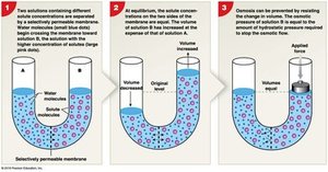
Diffusion is the movement of molecules from high to low concentration. Osmosis is the diffusion of water across a semi-permeable membrane in response to solute differences.
Simple diffusion: Small, nonpolar molecules (e.g., O2, CO2) cross the lipid bilayer.
Electrochemical gradient: For ions, movement is influenced by both chemical and electrical gradients.
Osmotic pressure: The force driving water movement into a solution.
Tonicity
Tonicity describes the effect of extracellular solutions on cell volume.
Isotonic: No net osmosis; cell volume unchanged.
Hypotonic: Net gain of water; cell may swell and burst (cytolysis).
Hypertonic: Net loss of water; cell shrinks (crenation).
Muscle Tissue and Structure
Types of Muscle Tissue
Muscle tissue is specialized for contraction and exists in three forms:
Skeletal muscle: Striated, voluntary, attached to bones.
Cardiac muscle: Striated, involuntary, found only in the heart.
Smooth muscle: Non-striated, involuntary, lines hollow organs.
Gross Anatomy of Skeletal Muscle
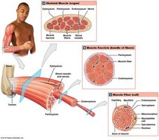
Skeletal muscles are organized into connective tissue layers that support and protect muscle fibers.
Endomysium: Surrounds individual muscle fibers.
Perimysium: Encloses bundles of fibers (fascicles).
Epimysium: Encloses the entire muscle.
Development and Structure of Skeletal Muscle Cells
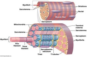
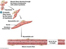
Skeletal muscle fibers are formed by the fusion of embryonic myoblasts, resulting in long, multinucleate cells. Satellite cells aid in muscle repair.
Microscopic Structure of Muscle Fibers
Muscle fibers contain myofibrils composed of myofilaments (actin and myosin). The sarcolemma is the cell membrane, and the sarcoplasmic reticulum stores calcium ions.
Myofilament Arrangement
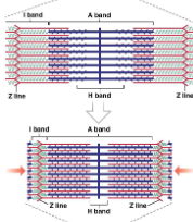
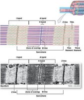
Thick (myosin) and thin (actin) filaments are organized into sarcomeres, the functional units of muscle contraction.
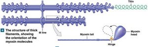
Thick filaments: Bundles of myosin molecules with heads that form cross-bridges during contraction.
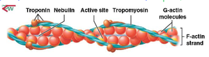
Thin filaments: Twisted strands of actin, with tropomyosin covering active sites and troponin regulating access.
Striated Sarcomeres
Sarcomeres are defined by Z lines and contain alternating bands:
I band: Light, only thin filaments.
A band: Dark, thick filaments and overlapping thin filaments.
H band: Only thick filaments.
Sliding Filament Model of Muscle Contraction
Sarcomere Structure and Function
Muscle contraction occurs as thin filaments slide over thick filaments, shortening the sarcomere and thus the muscle fiber.
Sliding Filament Theory
The sliding filament theory explains the steps of muscle contraction:
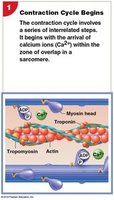
Contraction Cycle Begins: Calcium ions bind to troponin, exposing actin's active sites.
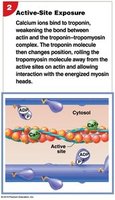
Active-Site Exposure: Troponin changes shape, moving tropomyosin and exposing actin's active sites.
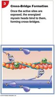
Cross-Bridge Formation: Energized myosin heads bind to exposed actin sites.
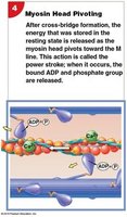
Myosin Head Pivoting (Power Stroke): Myosin head pivots, pulling actin toward the M line and releasing ADP and Pi.
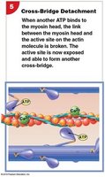
Cross-Bridge Detachment: ATP binds to myosin, causing it to detach from actin.
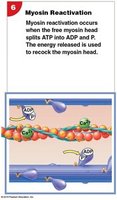
Myosin Reactivation: ATP is hydrolyzed, re-cocking the myosin head.
Role of ATP in Muscle Contraction
ATP provides energy for myosin head movement and is essential for both contraction and relaxation.
ATP hydrolysis:
Myosin heads bind ATP to detach from actin and re-cock for another cycle.
Muscle Contraction Mechanisms
Length-Tension Relationship
The amount of tension a muscle can produce depends on the overlap between actin and myosin filaments and the room for movement during contraction.
Frequency of Stimulation
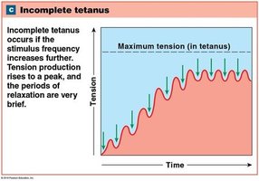
Muscle tension increases with repeated stimulation:
Twitch: Single, brief contraction.
Summation: Increased tension due to repeated stimulation.
Incomplete tetanus: Tension rises to a peak with brief relaxation periods.
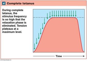
Complete tetanus: Tension plateaus at maximum level with no relaxation.
Motor Units and Tension Production
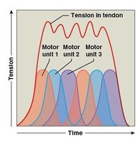
Motor units consist of a motor neuron and all the muscle fibers it innervates. Small motor units provide precise control, while large units provide gross movement.
Muscle Tone and Types of Contractions
Isotonic contraction: Muscle length changes as tension rises.
Isometric contraction: Tension rises but muscle length remains constant.
Energy Use and Muscle Contraction
ATP and Creatine Phosphate
Muscle cells store limited ATP and creatine phosphate (CP) for rapid energy release. Additional ATP is generated as needed.
Creatine phosphate reaction:
Aerobic and Anaerobic Metabolism
Aerobic respiration: Occurs in mitochondria, uses O2, produces CO2 and ATP.
Anaerobic metabolism: Produces ATP rapidly but inefficiently, leading to lactic acid buildup and fatigue.
Types of Skeletal Muscle Fibers
Slow, Fast, and Intermediate Fibers
Slow fibers (Type I): Small diameter, abundant mitochondria, fatigue-resistant.
Fast fibers (Type II-B): Large diameter, rapid contraction, fatigue quickly.
Intermediate fibers (Type II-A): Fast contraction, greater resistance to fatigue with training.
Muscle Fiber Distribution and Training Effects
Pale (white) muscles: Mostly fast fibers, suited for rapid, powerful contractions.
Dark (red) muscles: Dominated by slow fibers, suited for endurance activities.
Training increases intermediate fibers and fatigue resistance.
Muscle Hypertrophy and Atrophy
Hypertrophy: Increase in muscle diameter due to more actin and myosin.
Atrophy: Decrease in muscle diameter due to loss of myofilaments.
Muscle Disorders and Aging
Muscular System Disorders
Botulism: Blocks ACh release, causing paralysis.
Myasthenia gravis: Autoimmune disorder affecting ACh receptors.
Polio: Loss of motor neurons.
Multiple sclerosis: Damage to myelin, reducing action potential efficiency.
Tetanus: Excessive stimulation of motor neurons.
Aging and the Muscular System
Decrease in muscle fiber number and size
Reduced ATP, CP, glycogen, and myoglobin content
Increased fibrosis and decreased elasticity
Lower exercise tolerance and increased fatigue
Smooth and Cardiac Muscle
Smooth Muscle Structure and Function
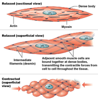
Smooth muscle is present in most organ systems and is non-striated. Thick filaments are scattered, and thin filaments attach to dense bodies.
Cardiac Muscle Structure and Function
Striated, involuntary, found only in the heart.
Cells linked by intercalated disks (desmosomes and gap junctions).
Automaticity: Can contract without neural stimulation.
Control of Contraction
Cardiac muscle: Controlled by pacemaker cells and autonomic nervous system.
Smooth muscle: Controlled by pacesetter cells, hormones, and autonomic nervous system.
Summary Table: Muscle Tissue Types
Feature | Skeletal Muscle | Cardiac Muscle | Smooth Muscle |
|---|---|---|---|
Striation | Striated | Striated | Non-striated |
Control | Voluntary | Involuntary | Involuntary |
Cell Structure | Multinucleate, long | Branched, single nucleus | Spindle-shaped, single nucleus |
Contraction | Tetanic | Single, rhythmic | Tetanic, sustained |
Energy Source | Aerobic/anaerobic | Aerobic | Aerobic/anaerobic |
