 Indietro
Microbiology Lab Techniques and Safety: Study Guide for Anatomy & Physiology Students
Guida di studio - Note intelligenti
 Appunti personalizzati basati sui tuoi materiali, ampliati con definizioni chiave, esempi e contesto.
Microbiology Laboratory Safety
General Safety Practices
Laboratory safety is essential for preventing accidents and ensuring a safe environment for all participants. Proper hygiene, attire, and awareness are fundamental.
Hand Hygiene: Wash hands thoroughly before and after handling lab materials, and after accidental contact with chemicals.
Attire: Wear closed-toed shoes, long pants, and tie back long hair. Avoid loose clothing and jewelry.
Food Restrictions: Do not eat, drink, or chew gum in the lab. Never taste chemicals.
Safety Equipment: Keep aisles clear, know the location of exits and safety equipment (fire extinguishers, blankets, showers, eyewash stations).
Personal Protective Equipment (PPE): Always wear safety glasses, gloves, and lab coats as required.
Documentation: Sign safety agreements, report injuries, and follow posted emergency procedures.
Chemistry and Microbiology Specific Safety
Chemistry: Wear PPE, avoid touching face, read chemical labels, dispose of chemicals properly, and exercise caution with burners and hot plates.
Microbiology: Disinfect benches, label biohazardous materials, use designated disposal, and separate note-taking from hazardous areas.
Microscope Structure and Use
Parts of a Microscope
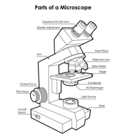
The microscope is a fundamental tool for observing microorganisms. Understanding its parts and their functions is crucial for accurate observation.
Eyepiece (Ocular lens): Usually 10x magnification; binocular for both eyes.
Objective lenses: Four lenses (4x, 10x, 40x, 100x) mounted on a nosepiece for varying magnification.
Stage: Holds the slide; mechanical stage allows precise movement.
Condenser: Focuses light on the specimen; includes an iris diaphragm for light adjustment.
Focus knobs: Coarse and fine adjustment for focusing the specimen.
Aseptic Technique and Streak Plate Method
Aseptic Technique
Aseptic technique is used to prevent contamination when culturing bacteria. It ensures pure cultures for study and safety.
Sterilization: Use autoclave or steam for equipment and media.
Flame sterilization: Sterilize loops and needles before transferring bacteria.
Flaming containers: Flame the mouth of tubes to prevent airborne contamination.
Streak Plate Technique
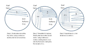
The streak plate method isolates individual bacterial colonies by diluting a sample across three sectors of a petri dish.
Procedure: Use a sterile loop to streak bacteria across three sectors, reflaming the loop between sectors.
Outcome: Individual colonies represent descendants of a single cell, allowing for pure culture isolation.
Microbial Ubiquity and Colony Morphology
Ubiquity of Microbes
Microorganisms are found everywhere in the environment, including extreme conditions. Their growth depends on nutrient availability, temperature, and atmospheric conditions.
Types: Bacteria, archaea, protozoa, algae, fungi, viruses, viroids, prions.
Role: Disease causation, nutrient cycling, decomposition.
Colony Morphology
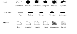
Microbial colonies can be classified by their form, elevation, and margin. These characteristics help identify and differentiate species.
Form: Punctiform, circular, filamentous, irregular, rhizoid, spindle.
Elevation: Flat, raised, convex, pulvinate, umbonate.
Margin: Entire, undulate, lobate, erose, filamentous, curled.
Staining Techniques
Simple and Negative Staining
Staining techniques enhance visualization of cells under the microscope. Simple stains color all cells, while negative stains color the background.
Simple Stain: Uses basic dyes (e.g., methylene blue) to stain cell structures.
Negative Stain: Uses acidic dyes (e.g., nigrosin) to stain the background, leaving cells clear.
Gram Stain
The Gram stain differentiates bacteria into Gram-positive and Gram-negative based on cell wall structure.
Procedure: Crystal violet (primary stain), iodine (mordant), alcohol (decolorizer), safranin (counterstain).
Result: Gram-positive bacteria retain crystal violet (purple); Gram-negative take up safranin (pink/red).
Special Structure Stains
Spore Stain: Malachite green stains endospores; safranin stains vegetative cells.
Capsule Stain: Crystal violet stains cells; copper sulfate washes excess dye, leaving capsules clear.
Flagella Stain: Leifson stain colors flagella for visualization.
Quantitative Microbiology: Serial Dilution and Viable Cell Count
Serial Dilution Method
Serial dilution is used to estimate the number of living microorganisms in a sample by counting colony forming units (CFU).
Procedure: Dilute sample in steps, plate aliquots, incubate, and count colonies.
Calculation: CFU/ml in original sample = Number of Colonies Counted × Dilution Factor × (ml of Dilution Plates)
Counting Range: Only plates with 30–300 colonies are considered accurate.
Antibiotics, Antiseptics, and Disinfectants
Kirby-Bauer Disk Diffusion Method
This method tests microbial sensitivity to antibiotics by measuring the zone of inhibition around antibiotic disks.
Procedure: Inoculate agar plate, place antibiotic disks, incubate, measure zone of inhibition.
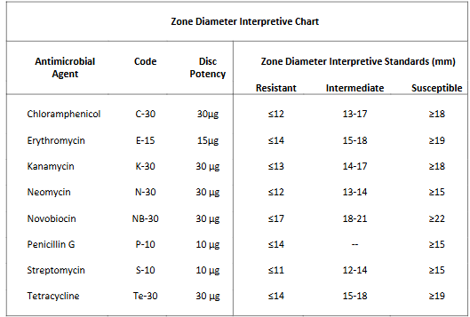
Interpretation: Compare zone diameter to standards to determine resistance, intermediate, or susceptibility.
Antimicrobial Agent | Code | Disc Potency | Resistant (mm) | Intermediate (mm) | Susceptible (mm) |
|---|---|---|---|---|---|
Chloramphenicol | C-30 | 30μg | ≤12 | 13–17 | ≥18 |
Erythromycin | E-15 | 15μg | ≤14 | 15–18 | ≥19 |
Kanamycin | K-30 | 30μg | ≤13 | 14–17 | ≥18 |
Neomycin | N-30 | 30μg | ≤12 | 13–14 | ≥15 |
Novobiocin | NB-30 | 30μg | ≤17 | 18–21 | ≥22 |
Penicillin G | P-10 | 10μg | ≤14 | -- | ≥15 |
Streptomycin | S-10 | 10μg | ≤11 | 12–14 | ≥15 |
Tetracycline | Te-30 | 30μg | ≤14 | 15–18 | ≥19 |
Biochemical Characterization of Gram Negative Bacteria
Fermentation Tubes
Used to test carbohydrate fermentation and gas production. Positive fermentation turns the indicator yellow and may produce gas in the Durham tube.
MR-VP Broth
MR Test: Detects mixed acid fermentation; positive result is a red color.
VP Test: Detects butanediol fermentation; positive result is a pink-burgundy color.
SIM Agar
Hydrogen Sulfide Production: Turns media black if positive.
Indole Production: Red color after adding Kovac’s reagent indicates positive.
Motility: Turbid growth away from stab line indicates motility.
Eosin Methylene Blue (EMB) Agar
Selective: Inhibits Gram-positive bacteria.
Differential: Lactose fermenters produce dark colonies; E. coli shows green sheen.
Example: E. coli on EMB agar produces dark colonies with a green sheen, indicating lactose fermentation.
