 Indietro
Motor Neurons and the Neuromuscular Junction: Mechanisms of Muscle Contraction
Guida di studio - Note intelligenti
 Appunti personalizzati basati sui tuoi materiali, ampliati con definizioni chiave, esempi e contesto.
Motor Neurons and the Neuromuscular Junction
Introduction
The neuromuscular junction is a specialized synapse where a motor neuron communicates with a skeletal muscle fiber, initiating muscle contraction. Understanding the anatomy and physiology of this junction is essential for comprehending how voluntary movements are controlled.
Ion Channels and Membrane Potentials
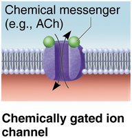
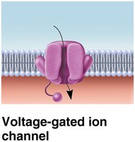
Ion channels are integral membrane proteins that regulate the flow of ions across the cell membrane, playing a crucial role in changing membrane potentials.
Chemically gated ion channels: Open in response to chemical messengers such as neurotransmitters (e.g., acetylcholine).
Voltage-gated ion channels: Open or close in response to changes in membrane potential.
Example: ACh receptors on muscle cells are chemically gated ion channels.
Anatomy of Motor Neurons and the Neuromuscular Junction
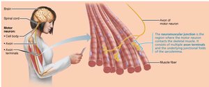
Skeletal muscles are stimulated by somatic motor neurons. The axons of these neurons extend from the central nervous system to the muscle, branching extensively as they approach the muscle fibers.
Axon terminals: The ends of axons form the neuromuscular junction (motor end plate) with muscle fibers.
Each muscle fiber is innervated by one motor neuron at a single neuromuscular junction.
Structure of the Neuromuscular Junction
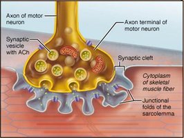
The neuromuscular junction consists of three main components:
Axon terminal: Contains synaptic vesicles filled with acetylcholine (ACh).
Synaptic cleft: A gel-filled space separating the axon terminal from the muscle fiber.
Junctional folds: Infoldings of the sarcolemma containing millions of ACh receptors.
Motor Unit and Recruitment
A motor unit consists of a motor neuron and all the muscle fibers it innervates. Recruitment refers to the activation of additional motor units to increase muscle contraction strength.
Motor unit: The functional unit for muscle contraction.
Recruitment: Allows graded control of muscle force.
Events Leading to Muscle Contraction
Overview of Muscle Contraction Steps
Four key steps must occur for skeletal muscle contraction:
Events at the neuromuscular junction
Muscle fiber excitation
Excitation-contraction coupling
Cross bridge cycling
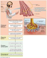
Events at the Neuromuscular Junction
The neuromuscular junction is the site where the motor neuron stimulates the muscle fiber, initiating the process of muscle contraction.
Action potential arrives at the axon terminal of the motor neuron.
Voltage-gated Ca2+ channels open, allowing Ca2+ to enter the axon terminal.
Ca2+ entry triggers the release of acetylcholine (ACh) by exocytosis.
ACh diffuses across the synaptic cleft and binds to receptors on the sarcolemma.
ACh binding opens ion channels, allowing Na+ to enter and K+ to exit, resulting in an end plate potential.
ACh is degraded by acetylcholinesterase, terminating its effects.
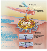
Generation of an Action Potential Across the Sarcolemma
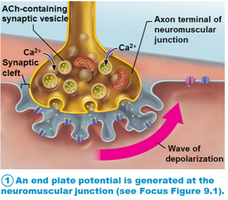
End Plate Potential
The binding of ACh to its receptors on the sarcolemma opens chemically gated ion channels, allowing Na+ influx and a small K+ efflux. This results in a local depolarization known as the end plate potential.
Depolarization
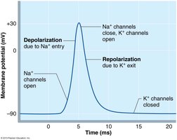
If the end plate potential reaches a critical threshold, voltage-gated Na+ channels open, causing a rapid influx of Na+ and generating an action potential. This action potential propagates along the sarcolemma, leading to muscle fiber contraction.
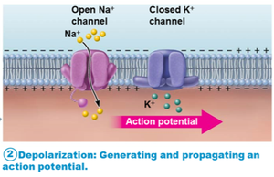
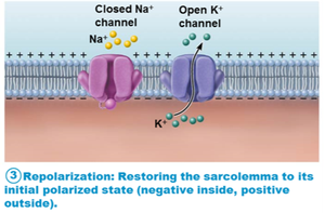
Repolarization
After depolarization, Na+ channels close and K+ channels open, allowing K+ to exit the cell. This restores the resting membrane potential. During the refractory period, the muscle fiber cannot be stimulated until repolarization is complete. The Na+-K+ pump restores ionic conditions.
Action Potential Tracing
Recording of an action potential in a muscle fiber shows the changes in membrane potential due to Na+ and K+ ion channel activity.
Excitation-Contraction Coupling
Mechanism of Excitation-Contraction Coupling
Excitation-contraction (E-C) coupling refers to the events that transmit the action potential along the sarcolemma and into the T tubules, leading to Ca2+ release from the sarcoplasmic reticulum (SR) and subsequent muscle contraction.
Action potential propagates along the sarcolemma and into T tubules.
Voltage-sensitive proteins in T tubules stimulate Ca2+ release from the SR.
Ca2+ release initiates contraction by enabling cross bridge cycling.
The action potential is brief and ends before contraction is visible.
Additional info: Cross bridge cycling involves the interaction of actin and myosin filaments, powered by ATP, resulting in muscle shortening.
