 Indietro
Movements and Axial Muscles: Anatomy & Physiology Lab Study Guide
Guida di studio - Note intelligenti
 Appunti personalizzati basati sui tuoi materiali, ampliati con definizioni chiave, esempi e contesto.
Movements of the Human Body
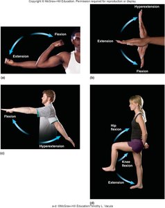
Flexion and Extension
Flexion and extension are fundamental movements that occur at many joints in the body. Flexion decreases the angle between two bones, while extension increases it. Hyperextension refers to extension beyond the normal range of motion.
Flexion: Bending a joint, such as the elbow or knee, so that the angle decreases.
Extension: Straightening a joint, increasing the angle between bones.
Hyperextension: Extension beyond anatomical position.
Example: Raising your arm forward (shoulder flexion), straightening your arm (elbow extension).
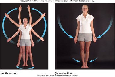
Abduction and Adduction
Abduction and adduction are movements along the frontal plane. Abduction moves a limb away from the midline, while adduction brings it toward the midline.
Abduction: Moving limbs laterally away from the body.
Adduction: Moving limbs medially toward the body.
Example: Raising arms sideways (abduction), lowering them to the sides (adduction).
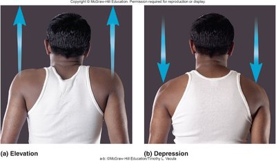
Elevation and Depression
Elevation and depression refer to raising or lowering a body part in the frontal plane.
Elevation: Lifting a body part superiorly (e.g., shrugging shoulders).
Depression: Moving a body part inferiorly (e.g., lowering shoulders).
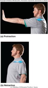
Protraction and Retraction
Protraction and retraction are movements along the transverse plane, often seen in the scapula or jaw.
Protraction: Moving a body part forward (anteriorly).
Retraction: Moving a body part backward (posteriorly).
Example: Pushing the jaw forward (protraction), pulling it back (retraction).
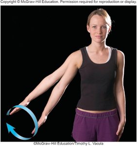
Circumduction
Circumduction is a circular movement that combines flexion, extension, abduction, and adduction. It is commonly seen in ball-and-socket joints like the shoulder.
Circumduction: Moving a limb in a circular motion.
Example: Drawing a circle with your arm.
Rotation
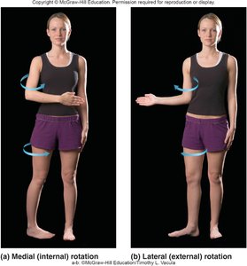
Rotation is the turning of a bone around its own longitudinal axis. It can be medial (internal) or lateral (external).
Medial (internal) rotation: Rotating a limb toward the midline.
Lateral (external) rotation: Rotating a limb away from the midline.
Example: Rotating the thigh inward or outward.
Supination and Pronation
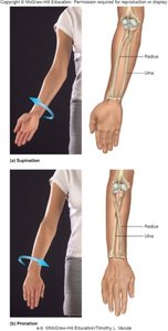
Supination and pronation are specific to the forearm. Supination turns the palm upward, while pronation turns it downward.
Supination: Rotating the forearm so the palm faces up.
Pronation: Rotating the forearm so the palm faces down.
Movements of the Head and Trunk
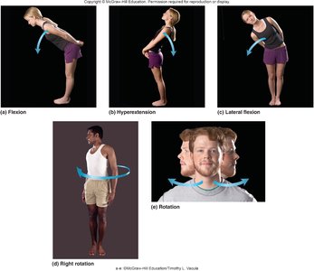
The head and trunk can perform flexion, extension, lateral flexion, and rotation. These movements are essential for posture and locomotion.
Flexion: Bending forward.
Extension: Bending backward.
Lateral flexion: Bending to the side.
Rotation: Turning the head or trunk.
Movements of the Hands and Digits
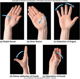
The hands and fingers exhibit specialized movements such as radial and ulnar flexion, abduction, palmar abduction, and opposition of the thumb.
Radial flexion: Moving the hand toward the thumb side.
Ulnar flexion: Moving the hand toward the little finger side.
Abduction of fingers: Spreading fingers apart.
Opposition of thumb: Touching the thumb to other fingers.
Movements of the Foot
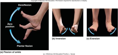
The foot can perform dorsiflexion, plantar flexion, inversion, and eversion. These movements are crucial for walking and balance.
Dorsiflexion: Raising the foot upward.
Plantar flexion: Pointing the foot downward.
Inversion: Turning the sole inward.
Eversion: Turning the sole outward.
Introduction to Muscles
Muscle Action and Terminology
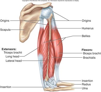
Muscles generate movement by contracting, which pulls on bones. They do not push. Key terms include origin (fixed attachment), insertion (mobile attachment), and belly (mid-portion).
Origin: The stationary attachment point of the muscle.
Insertion: The movable attachment point.
Belly: The thick, central part of the muscle.
Example: Biceps brachii originates from the scapula and inserts on the radius.
Muscle Classification by Fascicle Arrangement
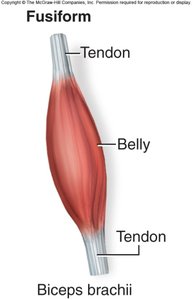
Muscles are classified based on the arrangement of their fascicles, which determines their strength and direction of pull.
Fusiform: Spindle-shaped, e.g., biceps brachii.
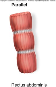
Parallel: Fascicles run parallel, e.g., rectus abdominis.
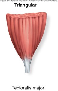
Convergent (Triangular): Fascicles converge to a single point, e.g., pectoralis major.
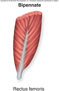
Pennate: Fascicles attach obliquely to a tendon, e.g., rectus femoris (bipennate).
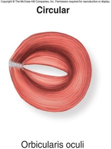
Circular: Fascicles arranged in rings, e.g., orbicularis oculi.
Muscle Roles: Agonist, Synergist, Antagonist
Muscles work together to produce movement. The agonist (prime mover) produces most of the force, the synergist aids the agonist, and the antagonist opposes the agonist.
Agonist: Main muscle responsible for movement.
Synergist: Assists the agonist and stabilizes the joint.
Antagonist: Opposes the agonist's action.
Example: Elbow flexion: brachialis (agonist), biceps brachii (synergist), triceps brachii (antagonist).
Innervation of Muscles
Muscles receive nerve supply from cranial or spinal nerves. Proper innervation is essential for muscle function.
Cranial nerves: I-XII, supply muscles of the head and neck.
Spinal nerves: Originate from the spinal cord, supply trunk and limb muscles.
Example: T1, S2, C3 indicate spinal nerve levels.
Axial Muscles: Overview and Functions
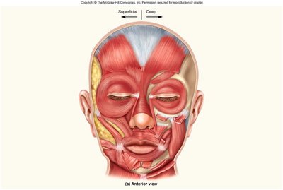
Muscles of Facial Expression
These muscles are responsible for facial movements and expressions. They are innervated by the Facial Nerve (CN VII).
Examples: Frontalis, orbicularis oculi, zygomaticus major/minor, risorius, buccinator, orbicularis oris, mentalis, platysma, nasalis.
Function: Express emotions, move facial skin.
Clinical note: Paralysis leads to sagging (e.g., stroke, Bell's palsy).
Muscles of Mastication (Mandible)
These muscles move the jaw for chewing and are innervated by the Trigeminal Nerve (CN V).
Examples: Temporalis, masseter, pterygoids.
Function: Elevate, depress, and move the mandible side-to-side.
Muscles of the Tongue
Muscles of the tongue are essential for speech and swallowing. They are innervated by the Hypoglossal Nerve (CN XII).
Examples: Styloglossus, genioglossus, hyoglossus.
Taste sensation: Anterior 2/3 by Facial Nerve (VII), posterior 1/3 by Glossopharyngeal Nerve (IX).
Muscles of the Pharynx and Hyoid
These muscles are important in swallowing and are innervated by CN IX and X.
Examples: Pharyngeal constrictors, stylohyoid.
Gross Head and Neck Movements
Muscles responsible for head and neck movements are innervated by the Accessory Nerve (CN XI) and various spinal nerves.
Examples: Sternocleidomastoid (SCM), trapezius, erector spinae.
Function: Flexion, extension, rotation, and lateral flexion of the head and neck.
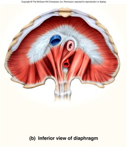
Muscles of Respiration
These muscles are essential for breathing and are innervated by the phrenic nerve and other spinal nerves.
Examples: External intercostals, internal intercostals, diaphragm.
Function: Expand and contract the thoracic cavity.
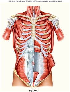
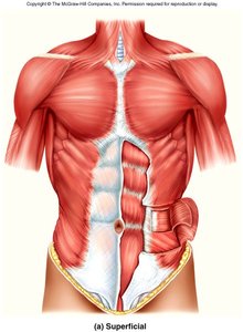
Muscles of the Abdomen
Abdominal muscles are innervated by T7-T12 spinal nerves and are involved in flexion, rotation, and regulation of intra-abdominal pressure.
Examples: Internal oblique, rectus abdominis, external oblique, transverse abdominal.
Function: Flex and rotate the torso, forceful exhalation, support abdominal organs.
Muscles of the Pelvis (Pelvic Floor Muscles)
Pelvic floor muscles support urogenital and digestive organs and are innervated by lumbar and sacral spinal nerves.
Examples: Levator ani, piriformis.
Function: Support pelvic organs, assist in continence.
Summary Table: Muscle Classification by Fascicle Arrangement
Type | Example Muscle | Function/Feature |
|---|---|---|
Fusiform | Biceps brachii | Spindle-shaped, strong contractions |
Parallel | Rectus abdominis | Uniform width, endurance |
Convergent (Triangular) | Pectoralis major | Broad origin, focused insertion |
Pennate (Bipennate) | Rectus femoris | Oblique fascicles, high force |
Circular | Orbicularis oculi | Ring-shaped, control openings |
Key Equations and Concepts
Muscle Force Generation
The force generated by a muscle depends on its cross-sectional area and the arrangement of its fascicles.
Equation: Muscle force is proportional to cross-sectional area:
CSA = cross-sectional area; specific tension = force per unit area
Practical Application
For lab assignments, students should be able to identify muscles, their functions (movements), and their innervation using models, diagrams, and photos.
Example: Frontalis muscle raises eyebrows, innervated by Facial Nerve (VII).
Additional info: Academic context was added to clarify muscle roles, innervation, and movement definitions.
