 Indietro
Muscle Contraction and Neuromuscular Junction: Study Notes
Guida di studio - Note intelligenti
 Appunti personalizzati basati sui tuoi materiali, ampliati con definizioni chiave, esempi e contesto.
Muscle Contraction: Sliding Filament Model
Overview of Muscle Contraction
Muscle contraction is the process by which muscle fibers generate force and shorten, enabling movement. The sliding filament model explains how actin and myosin filaments interact to produce contraction without changing their individual lengths.
Contraction: Activation of cross bridges (myosin heads binding to actin) generates force.
Shortening: Occurs when tension from cross bridges exceeds opposing forces.
Relaxation: Contraction ends when cross bridges become inactive.
Sliding Filament Model Details
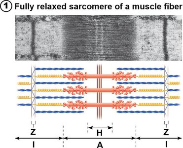
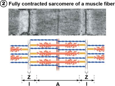
In the relaxed state, thin (actin) and thick (myosin) filaments overlap only slightly. During contraction, thin filaments slide past thick filaments, increasing overlap. Neither filament changes length; instead, the sarcomere shortens.
Cross Bridge Cycling: Myosin heads attach and detach from actin, pulling thin filaments toward the center of the sarcomere.
Sarcomere Changes: Z discs are pulled toward the M line, I bands shorten, H zone disappears, and A bands move closer together.
Example: The contraction of skeletal muscle during movement, such as lifting an object.
Nerve Stimulus and Events at the Neuromuscular Junction
Steps Required for Skeletal Muscle Contraction
For skeletal muscle to contract, four key steps must occur:
Nerve stimulation
Generation of an action potential in the sarcolemma
Propagation of the action potential along the sarcolemma
Brief rise in intracellular Ca2+ levels
Steps 1 and 2 occur at the neuromuscular junction (NMJ), while steps 3 and 4 are part of excitation-contraction coupling.
Structure of the Neuromuscular Junction
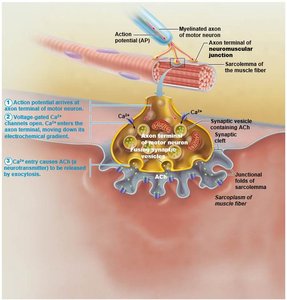
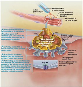
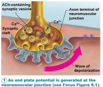
The NMJ is the site where a motor neuron communicates with a muscle fiber. It consists of the axon terminal, synaptic cleft, and junctional folds of the sarcolemma.
Axon Terminal: Contains synaptic vesicles filled with acetylcholine (ACh).
Synaptic Cleft: Gel-filled space separating the axon terminal and muscle fiber.
Junctional Folds: Infoldings of the sarcolemma with numerous ACh receptors.
Events at the Neuromuscular Junction
When a nerve impulse arrives at the axon terminal:
ACh is released into the synaptic cleft.
ACh binds to receptors on the sarcolemma, triggering electrical events that generate an action potential.
ACh is rapidly broken down by acetylcholinesterase, ending the signal.
Generation of an Action Potential Across the Sarcolemma
Resting Membrane Potential
The sarcolemma is polarized at rest, with the inside of the cell being negative relative to the outside. Action potentials are generated by changes in these electrical charges.
Steps in Action Potential Generation
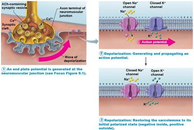
End Plate Potential: ACh binds to receptors, opening chemically gated ion channels. Na+ diffuses into the muscle fiber, making the interior less negative (depolarization).
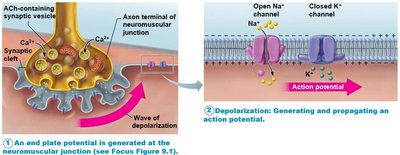
Depolarization: If the end plate potential reaches threshold, voltage-gated Na+ channels open, causing a large influx of Na+ and triggering an action potential. The AP propagates along the sarcolemma.
Repolarization: Na+ channels close, K+ channels open, and K+ exits the cell, restoring the resting membrane potential. The Na+/K+ pump reestablishes ionic conditions.
Membrane Potential Changes During Action Potential
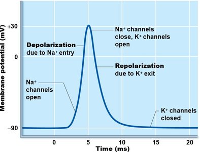
The action potential is characterized by rapid changes in membrane potential:
Depolarization: Na+ entry causes the membrane potential to become more positive.
Repolarization: K+ exit returns the membrane potential to its resting negative value.
Refractory Period: The muscle fiber cannot be stimulated again until repolarization is complete.
Key Equations
The ionic movements during action potential can be described by the Nernst equation for membrane potential:
Where E is the equilibrium potential, R is the gas constant, T is temperature, z is the ion charge, F is Faraday's constant, and the brackets denote ion concentration.
Summary Table: Events at the Neuromuscular Junction
Step | Description |
|---|---|
Nerve Impulse Arrival | Action potential arrives at axon terminal |
ACh Release | ACh released into synaptic cleft |
ACh Binding | ACh binds to sarcolemma receptors |
Action Potential Generation | Electrical events generate action potential |
ACh Breakdown | ACh broken down by acetylcholinesterase |
Example: The neuromuscular junction is essential for voluntary muscle contraction, such as moving the arm.
Additional info: Expanded explanations and context were added to ensure completeness and clarity for exam preparation.
