 Indietro
Muscle Contraction: Mechanisms and Regulation
Guida di studio - Note intelligenti
 Appunti personalizzati basati sui tuoi materiali, ampliati con definizioni chiave, esempi e contesto.
Muscle Contraction: Mechanisms and Regulation
Overview of Skeletal Muscle Contraction
Skeletal muscle contraction is a complex process involving electrical and chemical signals that result in the shortening of muscle fibers. Four essential steps must occur for contraction: events at the neuromuscular junction, muscle fiber excitation, excitation–contraction coupling, and cross-bridge cycling.
Events at the Neuromuscular Junction
Stepwise Activation of Muscle Fiber
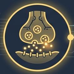
The neuromuscular junction (NMJ) is the site where a motor neuron communicates with a muscle fiber, triggering contraction. The process involves the release of neurotransmitter and subsequent depolarization of the muscle membrane.
Step 1: An action potential arrives at the axon terminal of the motor neuron.
Step 2: Voltage-gated Ca2+ channels open; Ca2+ enters the terminal.
Step 3: Ca2+ entry causes release of acetylcholine (ACh) into the synaptic cleft by exocytosis.
Step 4: ACh diffuses across the cleft and binds to ACh receptors on the sarcolemma.
Step 5: Receptor channels open: Na+ enters (more than K+ leaves) → end plate potential.
Step 6: Acetylcholinesterase degrades ACh, ending the signal.
Muscle Fiber Excitation
Generation and Propagation of Action Potential
Once the end plate potential is generated, it triggers an action potential that spreads across the sarcolemma, leading to muscle fiber excitation.
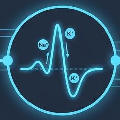
End Plate Potential: ACh opens chemically gated channels; Na+ diffuses in, causing local depolarization.
Depolarization: If threshold is reached, voltage-gated Na+ channels open; Na+ influx triggers an action potential (AP).
Repolarization: Na+ channels close and K+ channels open; K+ efflux restores the resting state. The refractory period prevents new stimulation until repolarization is complete.
Excitation–Contraction Coupling
Linking Electrical Signal to Calcium Release and Myofilament Sliding
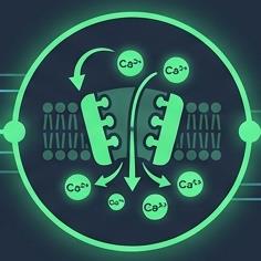
Excitation–contraction (E-C) coupling is the process by which the action potential on the sarcolemma leads to the release of Ca2+ from the sarcoplasmic reticulum (SR), enabling contraction.
Step 1: The AP propagates along the sarcolemma and down the T tubules.
Step 2: Voltage-sensitive T-tubule proteins change shape, opening SR Ca2+ release channels; Ca2+ floods the cytosol.
Step 3: Ca2+ binds troponin, which moves tropomyosin and exposes myosin-binding sites on actin.
Step 4: Myosin binds actin, forming cross bridges — contraction begins.
Aftermath: Ca2+ is pumped back into the SR; tropomyosin blocks again → relaxation.
Cross-Bridge Cycling
The Four-Step Molecular Cycle
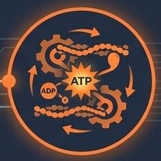
The cross-bridge cycle is the molecular mechanism by which actin filaments slide past myosin, resulting in muscle contraction. ATP is essential for this process.
Step 1: Cross-bridge formation: the high-energy myosin head attaches to an actin active site.
Step 2: Power stroke: ADP and Pi are released; the head pivots and pulls the thin filament toward the M line.
Step 3: Detachment: ATP binds myosin, weakening the bond; the cross bridge detaches.
Step 4: Cocking: ATP hydrolysis returns the head to its high-energy position — ready to bind again.
Example: Rigor mortis occurs when ATP is unavailable, preventing detachment of myosin heads.
Motor Units & Whole Muscle Contraction
Grading Muscle Force
The nervous system controls muscle force by varying the frequency and strength of stimulation. Motor units are recruited based on the size principle, and muscle contractions can be isotonic or isometric.
Motor Unit: A motor neuron and all the muscle fibers it supplies.
Muscle Twitch: The response of a muscle fiber to a single AP.
Graded Responses: Varying contraction strength by changing stimulus frequency (summation, tetanus) and stimulus strength (recruitment).
Isotonic Contraction: Muscle changes length and moves the load.
Isometric Contraction: Muscle tension rises, but the muscle neither shortens nor lengthens.
Energy & Fiber Types
ATP Regeneration and Muscle Fiber Classification
ATP is the sole energy source for muscle contraction and must be regenerated rapidly. Muscle fibers are classified based on their speed and metabolic pathway.
ATP Regeneration: Occurs via creatine phosphate, anaerobic glycolysis, and aerobic respiration.
Fiber Types: Slow oxidative, fast oxidative, and fast glycolytic fibers differ in speed, fatigue resistance, and metabolic pathway.
Smooth Muscle
Structure, Contraction, and Regulation
Smooth muscle fibers are spindle-shaped, lack striations, and are innervated by the autonomic nervous system. Contraction is slow and synchronized, relying on extracellular Ca2+ and calmodulin.
Structure: Spindle-shaped, single nucleus, no striations.
Innervation: Varicosities release neurotransmitter into diffuse junctions.
Calcium Handling: Most Ca2+ comes from outside the cell; caveolae facilitate rapid influx.
Contraction Mechanism: Ca2+ binds calmodulin, activating myosin kinase.
Regulation: Controlled by nerves, hormones, or local chemical changes.
Special Features: Stress-relaxation response, wide working range, and ability to contract over a large length range.
Skeletal vs. Smooth Muscle: Comparison Table
Feature | Skeletal Muscle | Smooth Muscle |
|---|---|---|
Fiber shape | Long, wide, striated; many nuclei | Spindle-shaped, short; one nucleus; no striations |
Innervation | Somatic motor neurons at NMJs | Autonomic varicosities at diffuse junctions |
Ca2+ source | SR (via T tubules) | Mostly extracellular (caveolae); some SR |
Ca2+ binds | Troponin | Calmodulin → myosin kinase |
Coupling | Each fiber stimulated individually | Gap junctions; some pacemaker cells |
Contraction | Fast; sliding filaments | Slow, synchronized, corkscrew; sliding filaments |
Key Takeaways
At the NMJ: AP → Ca2+ entry → ACh release → end plate potential; acetylcholinesterase ends the signal
The end plate potential triggers an AP that spreads across the sarcolemma and down the T tubules
E-C coupling: T-tubule proteins open SR channels; Ca2+ binds troponin, exposing myosin-binding sites
Cross-bridge cycle: attach → power stroke → detach (ATP) → cock (ATP hydrolysis)
Force is graded by stimulus frequency (summation, tetanus) and strength (recruitment, size principle)
ATP is regenerated by creatine phosphate, anaerobic glycolysis and aerobic respiration; fiber types differ in speed and pathway
Smooth muscle: extracellular Ca2+, calmodulin and myosin kinase; slow, synchronized, stretch-tolerant
