 Indietro
Muscle Contraction: Mechanisms and Types in Skeletal and Smooth Muscle
Guida di studio - Note intelligenti
 Appunti personalizzati basati sui tuoi materiali, ampliati con definizioni chiave, esempi e contesto.
Muscle Contraction: Overview
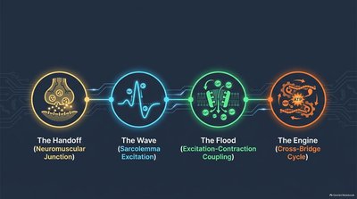
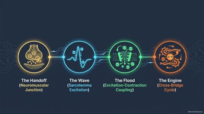
The Four Steps of Skeletal Muscle Contraction
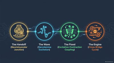
Skeletal muscle contraction is a complex process involving four sequential steps that ensure efficient movement and force generation. These steps are essential for understanding how muscles respond to neural stimulation and produce mechanical work.
Events at the Neuromuscular Junction: The initial communication between a motor neuron and a muscle fiber.
Muscle Fiber Excitation: The generation and propagation of an action potential across the muscle cell membrane.
Excitation-Contraction Coupling: The link between electrical signals and the release of calcium, leading to myofilament sliding.
Cross-Bridge Cycling: The molecular events that drive actin past myosin, resulting in contraction.
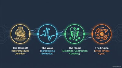
Events at the Neuromuscular Junction
Mechanism of Signal Transmission
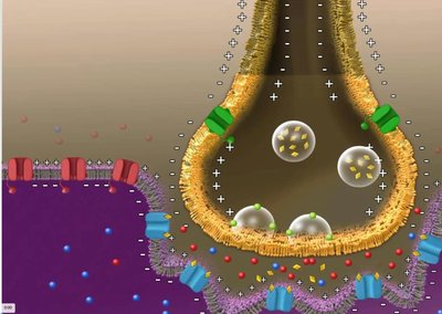
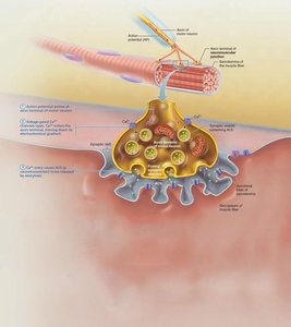
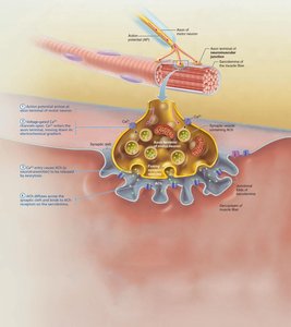
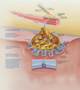
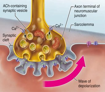
The neuromuscular junction (NMJ) is the site where a motor neuron communicates with a skeletal muscle fiber. This process involves the release of neurotransmitters and the initiation of muscle excitation.
Action Potential Arrival: An action potential reaches the axon terminal of the motor neuron.
Calcium Entry: Voltage-gated calcium channels open, allowing Ca2+ to enter the neuron.
Acetylcholine Release: Ca2+ triggers the release of acetylcholine (ACh) into the synaptic cleft.
ACh Binding: ACh binds to receptors on the sarcolemma, opening ion channels.
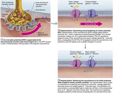
End Plate Potential: Na+ enters the muscle fiber, causing depolarization known as the end plate potential.
Acetylcholinesterase Action: The enzyme acetylcholinesterase degrades ACh, terminating the signal.
Muscle Fiber Excitation
Generation and Propagation of Action Potential
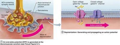
Once the muscle fiber is excited, an action potential is generated and propagated along the sarcolemma, leading to contraction.
End Plate Potential: ACh binding opens chemically gated ion channels, allowing Na+ influx and depolarization.
Depolarization: If threshold is reached, voltage-gated Na+ channels open, generating an action potential that spreads across the sarcolemma.
Repolarization: Na+ channels close, K+ channels open, and K+ efflux restores resting conditions. The refractory period prevents immediate re-stimulation.
Excitation–Contraction Coupling
Linking Electrical Signal to Mechanical Response
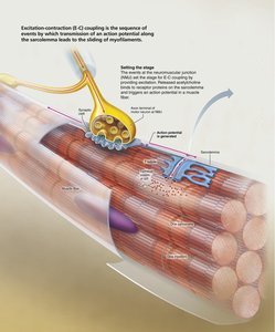
Excitation-contraction coupling is the process by which the action potential triggers calcium release, leading to the sliding of myofilaments and muscle contraction.
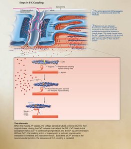
Action Potential Transmission: The action potential travels along the sarcolemma and into the T-tubules.
Calcium Release: Voltage-sensitive proteins trigger Ca2+ release from the sarcoplasmic reticulum (SR).
Myofilament Activation: Ca2+ binds to troponin, causing tropomyosin to move and expose actin binding sites.
Sliding Filament Mechanism: Myosin heads bind to actin, initiating contraction.
Cross-Bridge Cycling
Molecular Mechanism of Contraction
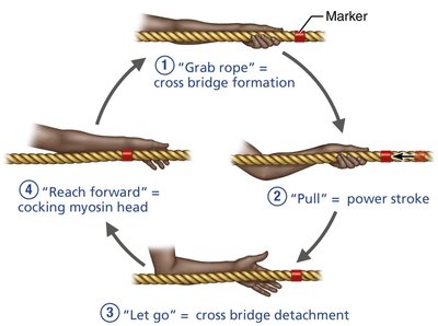
The cross-bridge cycle describes the repeated interactions between myosin and actin that produce muscle contraction. This cycle is analogous to pulling a rope with one hand.
Cross Bridge Formation: Myosin head attaches to actin active site.
Power Stroke: Myosin head pivots, pulling actin toward the M line.
Cross Bridge Detachment: ATP binds to myosin, causing detachment from actin.
Cocking of Myosin Head: ATP hydrolysis re-energizes the myosin head for the next cycle.
The Motor Unit and Types of Skeletal Muscle
Structure and Function of Motor Units
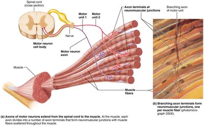
A motor unit consists of a motor neuron and all the muscle fibers it innervates. The number of fibers per motor unit determines the precision of muscle control.
Motor Unit: Functional unit composed of a motor neuron and its associated muscle fibers.
Fine Control: Smaller motor units allow for greater precision.
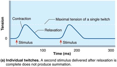
Muscle Twitch: The simplest contraction, resulting from a single action potential.
Muscle Twitch and Graded Responses
Phases and Variability of Muscle Twitch
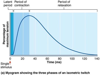
Muscle twitches consist of three phases and vary in strength and duration depending on muscle type and metabolic properties.
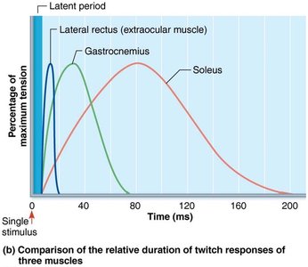
Latent Period: Time between stimulus and contraction; events of excitation-contraction coupling occur.
Period of Contraction: Cross bridge formation and tension increase.
Period of Relaxation: Ca2+ reentry into SR; tension declines.
Variability: Eye muscles contract rapidly and briefly; larger muscles contract more slowly and sustain tension longer.
Graded Muscle Responses
Modulation of Muscle Contraction
Muscle contraction strength is modulated by changing the frequency and strength of stimulation. This allows for precise control of skeletal movement.
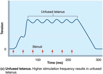
Wave (Temporal) Summation: Increased frequency of stimulation leads to summation of contractions.
Unfused (Incomplete) Tetanus: Higher frequency results in sustained, quivering contractions.
Fused (Complete) Tetanus: Maximum tension is reached; contractions fuse into a smooth plateau.
Recruitment: Increasing stimulus strength activates more motor units, enhancing contractile force.
Size Principle: Smaller motor units are recruited first, followed by larger units for powerful contractions.
Types of Muscle Contractions
Isotonic and Isometric Contractions
Muscle contractions can be classified based on whether the muscle changes length or not during contraction.
Isotonic Contractions: Muscle changes length and moves the load. Includes concentric (muscle shortens) and eccentric (muscle lengthens) contractions.
Isometric Contractions: Muscle tension increases, but length remains unchanged; occurs when load exceeds muscle's capacity.
Energy for Muscle Contraction
ATP Regeneration Pathways
ATP is the sole energy source for muscle contraction and must be regenerated rapidly through three main pathways.
Direct Phosphorylation: Creatine phosphate (CP) donates a phosphate to ADP to form ATP.
Anaerobic Pathway: Glycolysis and lactic acid formation provide ATP without oxygen.
Aerobic Pathway: Oxidative phosphorylation produces ATP using oxygen.
Pathway | Substrate | ATP Yield | Duration |
|---|---|---|---|
Direct Phosphorylation | Creatine Phosphate | 1 ATP per CP | 15 seconds |
Anaerobic Glycolysis | Glucose | 2 ATP per glucose | 30-40 seconds |
Aerobic Respiration | Glucose, fatty acids | ~32 ATP per glucose | Hours |
Aerobic Endurance: Duration a muscle can contract using aerobic pathways. Anaerobic Threshold: Point where metabolism shifts to anaerobic pathway.
Muscle Fiber Types
Classification and Functional Properties
Muscle fibers are classified based on contraction speed and metabolic pathway used for ATP synthesis.
Slow Fibers: Slow contraction, high endurance, use aerobic pathways.
Fast Fibers: Rapid contraction, fatigue quickly, use anaerobic glycolysis.
Oxidative Fibers: Rely on aerobic metabolism.
Glycolytic Fibers: Rely on anaerobic glycolysis.
Most muscles contain a mixture of fiber types, and genetics determine individual proportions.
Smooth Muscle
Structural and Functional Differences from Skeletal Muscle
Smooth muscle fibers are spindle-shaped, shorter, and have a single nucleus. They lack striations and connective tissue sheaths, and are innervated by the autonomic nervous system.
Varicosities: Bulbous nerve fiber swellings release neurotransmitters into diffuse junctions.
Sarcoplasmic Reticulum: Less elaborate, most Ca2+ comes from extracellular space.
Gap Junctions: Electrically connect cells for synchronized contraction.
Myofilament Arrangement: Thick and thin filaments arranged diagonally; contraction is corkscrew-like.
No Troponin: Ca2+ binds to calmodulin instead.
Dense Bodies: Anchor filaments to sarcolemma, analogous to Z discs.
Mechanism and Regulation of Smooth Muscle Contraction
Smooth muscle contracts slowly and synchronously, with some cells acting as pacemakers. Contraction is triggered by increased intracellular Ca2+, which binds to calmodulin and activates myosin kinase, leading to cross-bridge formation.
Relaxation: Requires Ca2+ detachment, active transport, and dephosphorylation of myosin.
Regulation: Controlled by nerves, hormones, or local chemical changes.
Special Features: Stress-relaxation response, ability to contract over a wide range of lengths, and adaptation to new lengths.
Example: The stomach and bladder can store contents temporarily due to smooth muscle's ability to adapt to stretch.
