 Indietro
Muscle Contraction, Metabolism, and Fiber Types: Study Notes for Anatomy & Physiology
Guida di studio - Note intelligenti
 Appunti personalizzati basati sui tuoi materiali, ampliati con definizioni chiave, esempi e contesto.
Contraction of Skeletal Muscle
Muscle Mechanics & Principles
Skeletal muscle contraction is governed by several mechanical principles that determine force generation and movement. Understanding these principles is essential for comprehending how muscles produce tension and respond to loads.
Muscle Tension: The force exerted by a contracting muscle.
Load: The opposing force due to the weight of the object the muscle is pulling against.
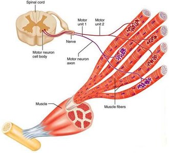
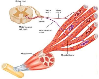
Motor Unit: A motor neuron and all the muscle fibers it innervates. When the motor neuron fires, all fibers it innervates contract simultaneously. Motor units may contain few fibers (for fine control) or many fibers (for powerful movements), and fibers are scattered throughout the muscle.
Muscle Twitch
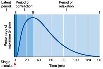
A muscle twitch is the response of a motor unit to a single action potential from a motor neuron. It consists of three distinct phases:
Latent Period: Time between stimulus and onset of contraction.
Contraction Period: Muscle fibers shorten and tension increases.
Relaxation Period: Muscle tension decreases as fibers return to resting state.
Graded Muscle Responses
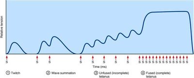
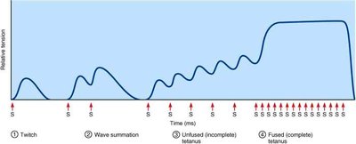
Graded responses allow muscles to produce smooth, coordinated contractions with varying strength and duration. These responses are achieved through:
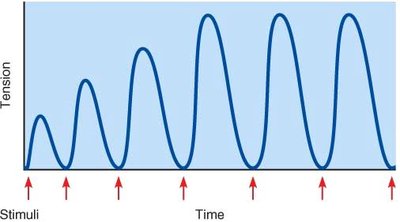
Wave Summation: Rapid, repeated stimuli result in stronger contractions due to incomplete relaxation and residual Ca2+ in the cytosol.
Unfused (Incomplete) Tetanus: Quivering contraction with partial relaxation between stimuli.
Fused (Complete) Tetanus: Smooth, sustained contraction with no relaxation between stimuli.
Recruitment
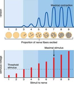
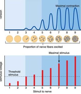
Recruitment refers to the activation of additional motor units to increase the force of contraction. The size principle states that:
Weak stimulus excites few small motor units.
Strong stimulus excites more and larger motor units.
Motor units are typically activated asynchronously to allow for sustained contractions.
Treppen (Staircase Effect)
Treppen describes the phenomenon where initial contractions are weaker, but subsequent contractions become stronger as the muscle "warms up." This is due to increased Ca2+ availability and enzyme activity.
Isotonic & Isometric Contractions
Muscle contractions can be classified based on whether the muscle changes length or not:
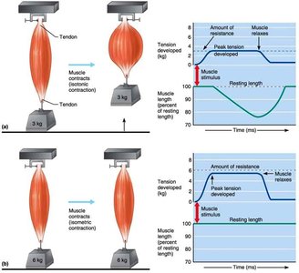
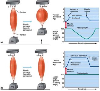
Isotonic Contraction: Muscle changes length while tension remains constant. Includes:
Concentric Contraction: Muscle shortens as it generates force.
Eccentric Contraction: Muscle lengthens while generating force.
Isometric Contraction: Muscle generates force without changing length.
Muscle Tone
Muscle tone is the state of partial contraction maintained by spinal reflexes. It keeps muscles ready for action, maintains posture, and stabilizes joints.
Muscle Metabolism
Energy Sources for Contraction (ATP)
Muscle contraction requires ATP, which is supplied by several mechanisms:
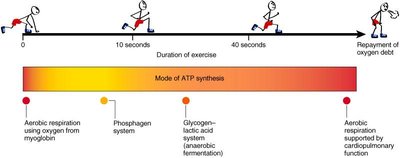
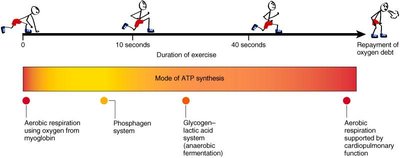
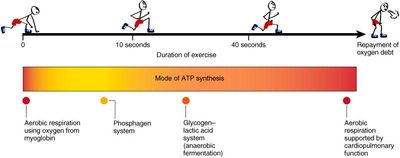
Stored ATP: Limited supply (~5 seconds), supplemented by O2 from myoglobin.
Phosphogen System: Rapid ATP production by transferring phosphate from creatine phosphate to ADP. Provides energy for ~15 seconds.
Anaerobic Respiration: Glycogen breakdown to lactic acid. Supplies energy for 30–40 seconds, but yields less ATP and causes lactic acid buildup.
Aerobic Respiration: Glucose breakdown in the presence of O2. Provides sustained energy for minutes to hours, with high ATP yield.
Muscle Fatigue
Muscle fatigue is the physiological inability to contract after prolonged activity. Causes include:
Decline in ATP, slowing enzyme activity.
Ionic disturbances, such as excess K+ in extracellular fluid and T-tubules.
Lactic acid buildup, lowering sarcoplasmic pH.
Quick fatigue leads to quick recovery, while slow-developing fatigue requires longer recovery.
Oxygen Debt
Oxygen debt is the extra O2 required after exercise to replenish the phosphogen system and anaerobic sources, convert lactic acid to pyruvic acid, and restore glycogen. It is manifested as heavy breathing after exercise.
Force of Muscle Contraction
Determinants of Contractile Force
The force generated by a muscle depends on several factors:
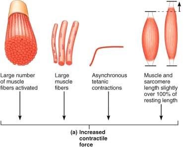
Number of fibers stimulated: More fibers activated increases force.
Size of fibers stimulated: Larger fibers generate more force.
Frequency of stimulation: Higher frequency increases force.
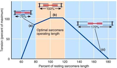
Degree of muscle stretch (pre-load): Muscles are positioned near optimal pre-stretch range (80–120% of optimal length) for maximal force.
Muscle Fiber Types
Classification of Skeletal Muscle Fibers
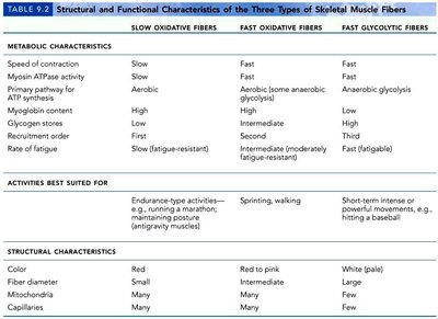
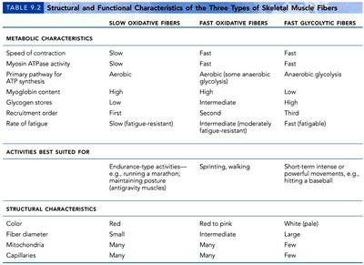
Skeletal muscle fibers are classified based on their metabolic and structural characteristics:
Characteristic | Slow Oxidative Fibers | Fast Oxidative Fibers | Fast Glycolytic Fibers |
|---|---|---|---|
Speed of contraction | Slow | Fast | Fast |
Myosin ATPase activity | Slow | Fast | Fast |
Primary pathway for ATP synthesis | Aerobic | Aerobic (some anaerobic) | Anaerobic glycolysis |
Myoglobin content | High | High | Low |
Glycogen stores | Low | Intermediate | High |
Recruitment order | First | Second | Third |
Rate of fatigue | Slow (fatigue-resistant) | Intermediate | Fast (fatigable) |
Activities best suited for | Endurance (marathon) | Sprinting, walking | Short-term intense (baseball) |
Color | Red | Red to pink | White |
Fiber diameter | Small | Intermediate | Large |
Mitochondria | Many | Many | Few |
Capillaries | Many | Many | Few |
Effects of Exercise
Muscle Adaptation and Atrophy
Muscles adapt to increased activity by becoming stronger and more efficient. Inactivity leads to atrophy, characterized by weakness and wasting away.
Aerobic Exercise: Activities utilizing aerobic respiration, such as running or swimming, improve endurance and cardiovascular health.
Anaerobic (Resistance) Exercise: Activities involving contractions against resistance, such as weightlifting, increase muscle mass and strength.
Disuse Atrophy: Muscle wasting due to inactivity.
Smooth Muscle
Structure (Cellular Level)
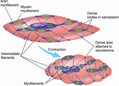
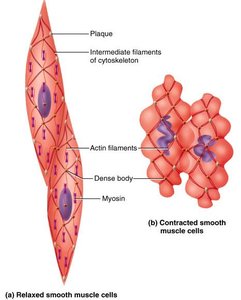
Smooth muscle cells are spindle-shaped, with thick and thin filaments interdigitating but lacking sarcomeres. Dense bodies serve as anchoring points for thin filaments and transmit force to connective tissue outside the cells.
Innervation
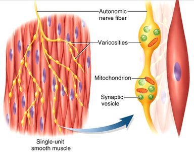
Smooth muscle is primarily innervated by the autonomic nervous system. Diffuse junctions, or varicosities, are bulbous swellings of autonomic nerves that release neurotransmitters.
Smooth Muscle Contraction (Organ Level)
Smooth muscle cells are often connected in sheets and linked by gap junctions, allowing rapid signaling and coordinated contraction. Contraction has a slower onset and longer duration compared to skeletal muscle.
E-C Coupling in Smooth Muscle
Excitation-contraction (E-C) coupling in smooth muscle involves:
Ca2+ enters cytosol.
Ca2+ binds and activates calmodulin.
Calmodulin activates myosin light chain kinase (MLCK).
MLCK phosphorylates myosin head group.
Activated myosin cross-bridges with actin filaments.
As Ca2+ levels decrease, myosin light chain phosphatase (MLCP) dephosphorylates myosin, inactivating it.
Stimulation of Smooth Muscle Contraction
Smooth muscle contraction can be stimulated by:
Neural stimuli (e.g., acetylcholine, norepinephrine)
Hormones
Histamine
Low O2, high CO2, low pH
Single-Unit (Visceral) Smooth Muscle
Single-unit smooth muscle is found in the walls of hollow organs. Cells are electrically coupled by gap junctions and contract rhythmically as a unit, often with spontaneous action potentials generated by pacemaker cells.
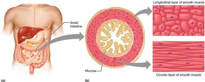
Multi-Unit Smooth Muscle
Multi-unit smooth muscle fibers are structurally independent, with many nerve endings for innervation. Responsiveness is involuntary, and these fibers are found in large airways, arteries, and internal eye muscles.
Additional Aspects of Muscles
Sex Differences and Aging
Testosterone: Favors skeletal muscle development; males have ~42% muscle body mass, females ~36%.
Sarcopenia: Gradual loss of muscle mass after age 30, which can be minimized by exercise, especially resistance training.
