 Indietro
Muscle Fiber Contraction: Excitation-Contraction Coupling and Cross Bridge Cycling
Guida di studio - Note intelligenti
 Appunti personalizzati basati sui tuoi materiali, ampliati con definizioni chiave, esempi e contesto.
Muscle Fiber Contraction
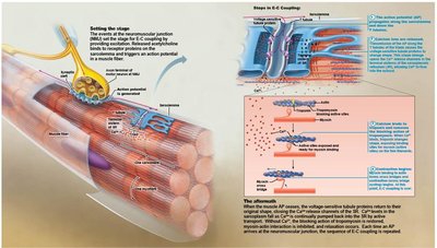
Excitation-Contraction (E-C) Coupling
Excitation-contraction (E-C) coupling refers to the sequence of events that link the electrical stimulus (action potential) of a muscle fiber to its mechanical contraction. This process ensures that the signal from the nervous system is efficiently translated into muscle movement.
Action Potential (AP) Propagation: The AP travels along the sarcolemma and into the T tubules.
Voltage-sensitive Proteins: These proteins in the T tubules respond to the AP and trigger the release of calcium ions (Ca2+) from the sarcoplasmic reticulum (SR).
Calcium Release: The influx of Ca2+ into the cytosol initiates muscle contraction.
Brief AP: The AP is short-lived and ends before the contraction is visible.
Role of Calcium in Muscle Contraction
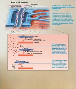
Calcium ions play a critical regulatory role in muscle contraction. Their concentration within the muscle fiber determines whether contraction can occur.
Low Intracellular Ca2+: Tropomyosin blocks the active sites on actin, preventing myosin from binding. The muscle fiber remains relaxed.
High Intracellular Ca2+: Ca2+ binds to troponin, causing a conformational change that moves tropomyosin away from the myosin-binding sites on actin. This allows myosin heads to bind to actin, forming cross bridges and initiating contraction.
Termination of Contraction: When nervous stimulation ceases, Ca2+ is pumped back into the SR, ending contraction.
Cross Bridge Cycling
Mechanism of Cross Bridge Cycling
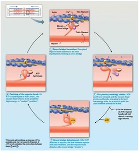
Cross bridge cycling is the fundamental process by which muscle fibers contract. It involves repeated interactions between myosin heads and actin filaments, powered by ATP.
Step 1: Cross Bridge Formation
High-energy myosin head binds to exposed actin site, forming a cross bridge.
Step 2: Power Stroke
Myosin head pivots, pulling actin toward the M line. This movement is called the power stroke.
Step 3: Cross Bridge Detachment
ATP binds to myosin head, causing it to detach from actin.
Step 4: Cocking of Myosin Head
ATP is hydrolyzed to ADP and inorganic phosphate, re-energizing ("cocking") the myosin head for the next cycle.
Summary Table: Steps in Cross Bridge Cycle
Step | Description |
|---|---|
1. Cross Bridge Formation | Myosin head binds to actin |
2. Power Stroke | Myosin head pivots, pulling actin |
3. Cross Bridge Detachment | ATP binds to myosin, causing detachment |
4. Cocking of Myosin Head | ATP hydrolysis re-energizes myosin head |
Key Terms and Concepts
Sarcolemma: The plasma membrane of a muscle fiber.
T tubules: Invaginations of the sarcolemma that help transmit the AP deep into the muscle fiber.
Sarcoplasmic Reticulum (SR): Specialized endoplasmic reticulum that stores and releases Ca2+.
Troponin and Tropomyosin: Regulatory proteins that control access to actin's myosin-binding sites.
ATP: The energy molecule required for both detachment and re-cocking of the myosin head.
Relevant Equations
ATP Hydrolysis:
Example: Application in Muscle Physiology
During activities such as running or lifting weights, the nervous system sends repeated action potentials to muscle fibers, triggering E-C coupling and cross bridge cycling. The rapid cycling of myosin heads and actin filaments results in sustained muscle contraction and force generation.
