 Indietro
Muscle Identification and Function Review for Anatomy & Physiology
Guida di studio - Note intelligenti
 Appunti personalizzati basati sui tuoi materiali, ampliati con definizioni chiave, esempi e contesto.
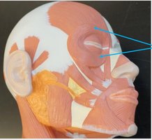
Q1. Identify the muscle indicated in the image and describe its function.
Background
Topic: Muscles of Facial Expression
This question tests your ability to recognize facial muscles and understand their actions, which is essential for learning about the musculoskeletal system in Anatomy & Physiology.
Key Terms:
Orbicularis oculi: A circular muscle around the eye.
Action: Closes the eyelids and allows for blinking and squinting.
Step-by-Step Guidance
Observe the location of the muscle indicated by the arrows. It surrounds the eye socket.
Recall that the muscle encircling the eye is responsible for closing the eyelids.
Think about the function: This muscle contracts to squeeze the eye shut, which is important for protecting the eye and spreading tears.
Review the name of the muscle that performs this action and its anatomical position.
Try solving on your own before revealing the answer!
Final Answer: Orbicularis oculi
The muscle indicated is the orbicularis oculi. Its function is to close the eye and squeeze the eye shut, which is essential for blinking and protecting the eye.
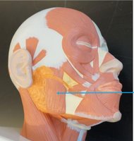
Q2. Identify the muscle indicated in the image and describe its function.
Background
Topic: Muscles of Mastication (Chewing)
This question focuses on the muscles involved in moving the jaw, which are critical for chewing and speaking.
Key Terms:
Masseter: A thick muscle located at the jaw.
Action: Elevates the mandible (jawbone) to close the mouth.
Step-by-Step Guidance
Look at the muscle indicated by the arrow, which is located at the side of the jaw.
Recall which muscle is responsible for elevating the mandible during chewing.
Consider the anatomical position and thickness of the muscle in this region.
Review the name and primary function of this muscle.
Try solving on your own before revealing the answer!
Final Answer: Masseter
The muscle indicated is the masseter. Its function is to elevate the mandible, allowing you to close your mouth and chew food.
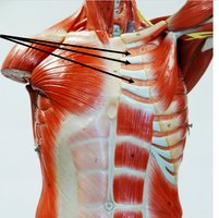
Q3. Identify the muscle indicated in the image and describe its function.
Background
Topic: Muscles of Respiration
This question tests your knowledge of the muscles between the ribs that are involved in breathing.
Key Terms:
Intercostal muscles: Muscles located between the ribs.
Action: Pulls ribs inferiorly to cause expiration (exhalation).
Step-by-Step Guidance
Examine the arrows pointing to the muscles between the ribs.
Recall the two types of intercostal muscles: internal and external.
Think about which type is responsible for pulling the ribs downward during exhalation.
Review the function of these muscles in the respiratory process.
Try solving on your own before revealing the answer!
Final Answer: Internal intercostals
The muscles indicated are the internal intercostals. Their function is to pull the ribs inferiorly, aiding in expiration (exhalation).
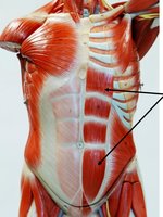
Q4. Identify the muscle indicated in the image and describe its function.
Background
Topic: Muscles of the Abdominal Wall
This question focuses on the muscle responsible for flexing the torso at the vertebral column.
Key Terms:
Rectus abdominis: A long, flat muscle running vertically along the front of the abdomen.
Action: Flexion of the torso at the vertebral column.
Step-by-Step Guidance
Observe the muscle indicated by the arrows, which is located in the midline of the abdomen.
Recall which muscle is responsible for movements such as sit-ups and bending forward.
Think about the anatomical features of this muscle, including its segmented appearance.
Review the name and primary function of this muscle.
Try solving on your own before revealing the answer!
Final Answer: Rectus abdominis
The muscle indicated is the rectus abdominis. Its function is to flex the torso at the vertebral column, allowing movements like bending forward.
Q5. Identify the muscle indicated in the image and describe its function.
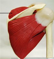
Background
Topic: Muscles of the Shoulder (Rotator Cuff)
This question tests your knowledge of the muscles that stabilize and move the shoulder joint.
Key Terms:
Supraspinatus: A muscle located above the spine of the scapula.
Action: Abduction of the arm at the shoulder.
Step-by-Step Guidance
Examine the muscle indicated by the arrow, which is located on the upper part of the scapula.
Recall the rotator cuff muscles and their functions.
Think about which muscle initiates abduction of the arm.
Review the name and primary function of this muscle.
Try solving on your own before revealing the answer!
Final Answer: Supraspinatus
The muscle indicated is the supraspinatus. Its function is to abduct the arm at the shoulder, especially the first 15 degrees of movement.
Q6. Identify the muscle indicated in the image and describe its function.
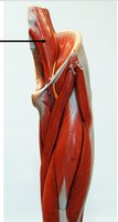
Background
Topic: Muscles of the Thigh
This question focuses on the muscles responsible for extension of the leg at the knee.
Key Terms:
Vastus lateralis: A muscle located on the lateral side of the thigh.
Action: Extension of the leg at the knee.
Step-by-Step Guidance
Observe the muscle indicated by the arrow, which is on the outer side of the thigh.
Recall the quadriceps group and their functions.
Think about which muscle is responsible for extending the knee.
Review the name and primary function of this muscle.
Try solving on your own before revealing the answer!
Final Answer: Vastus lateralis
The muscle indicated is the vastus lateralis. Its function is to extend the leg at the knee, which is important for walking, running, and jumping.
Q7. Identify the muscle indicated in the image and describe its function.
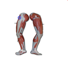
Background
Topic: Muscles of the Hip and Thigh
This question tests your knowledge of the muscles involved in abduction and stabilization of the hip.
Key Terms:
Tensor fasciae latae: A muscle located on the lateral side of the thigh.
Action: Abduction and medial rotation of the thigh; stabilizes the hip.
Step-by-Step Guidance
Examine the muscle indicated by the circle, which is on the lateral aspect of the thigh.
Recall the muscles that contribute to hip abduction and stabilization.
Think about the anatomical location and function of this muscle.
Review the name and primary function of this muscle.
Try solving on your own before revealing the answer!
Final Answer: Tensor fasciae latae
The muscle indicated is the tensor fasciae latae. Its function is to abduct and medially rotate the thigh, as well as stabilize the hip.
