 Indietro
Muscle Physiology and Cardiac Function: Structured Study Notes
Guida di studio - Note intelligenti
 Appunti personalizzati basati sui tuoi materiali, ampliati con definizioni chiave, esempi e contesto.
Muscle Physiology
Organization of Skeletal Muscle
Skeletal muscle is organized in a hierarchical structure, allowing efficient force generation and control. The muscle is composed of bundles called fascicles, which contain muscle fibers, each packed with myofibrils made of myofilaments.
Epimysium: Surrounds the whole muscle.
Perimysium: Surrounds fascicles.
Endomysium: Surrounds individual muscle fibers.
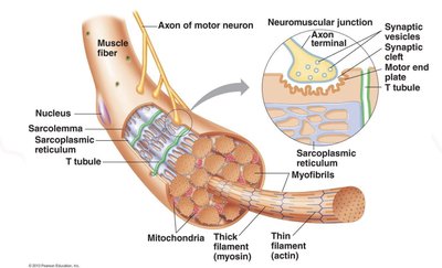
Skeletal muscle fibers: Long, cylindrical, striated cells with multiple nuclei.
Sarcolemma: Muscle cell membrane.
Sarcoplasm: Muscle cell cytoplasm.
Myofibrils: Contractile structures containing overlapping actin (thin) and myosin (thick) filaments.
Key Structures Inside a Muscle Fiber
Muscle fibers contain specialized structures for contraction and signal transmission.
T-tubules: Inward extensions of the sarcolemma that carry electrical signals into the cell.
Sarcoplasmic reticulum (SR): Stores and releases calcium to initiate contraction.
Triad: One T-tubule and two terminal cisternae of the SR.
Sarcomere: The Functional Unit
The sarcomere is the basic contractile unit of muscle, defined by Z discs.
Z disc: Boundary of a sarcomere.
M line: Center of the sarcomere, anchoring myosin filaments.
A band: Full length of myosin filaments; remains constant during contraction.
I band: Region with actin but no myosin; shortens during contraction.
H zone: Central region with only myosin; decreases during contraction.
Regulatory and Structural Proteins
Muscle contraction is regulated by proteins that control access to binding sites and provide structural support.
Tropomyosin: Covers actin binding sites in relaxed muscle.
Troponin: Binds calcium, moves tropomyosin, exposes binding sites.
Titin: Elastic protein anchoring myosin and supporting recoil.
Myosin heads: Contain actin-binding and ATPase sites.
Sliding Filament Mechanism
Muscle contraction occurs as actin filaments slide past myosin, shortening the sarcomere without changing filament length.
Calcium: Exposes actin binding sites for myosin attachment.
Power stroke: Myosin pulls actin toward the M line.
Band changes: A band stays constant; I band and H zone decrease.
The Crossbridge Cycle & ATP
The crossbridge cycle describes the steps of muscle contraction and the role of ATP.
Calcium exposes actin binding sites.
Energized myosin heads bind to actin.
Phosphate release drives the power stroke.
ATP binds to myosin, causing detachment from actin.
ATP hydrolysis re-energizes myosin for another cycle.
Rigor mortis: Occurs when ATP is unavailable, preventing crossbridge detachment.
Neuromuscular Junction (NMJ) and Synaptic Transmission
The NMJ is the site where motor neurons stimulate muscle fibers, initiating contraction.
Somatic motor neurons release acetylcholine (ACh) into the synaptic cleft.
ACh binds to receptors on the motor end plate, depolarizing the sarcolemma.
End-Plate Potential (EPP) triggers an action potential, propagating across the sarcolemma and down T-tubules.
Excitation-Contraction Coupling & Calcium Gating
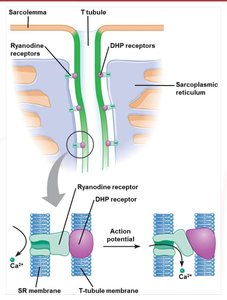
Excitation-contraction coupling links the electrical signal from the NMJ to muscle contraction via calcium release.
Action potentials activate DHP receptors in T-tubules.
DHP receptors mechanically open ryanodine receptors in the SR.
Calcium is released into the sarcoplasm, binding to troponin and enabling contraction.
The Motor Unit & Muscle Twitch
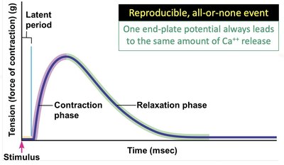
A motor unit consists of a single motor neuron and all the muscle fibers it innervates. Muscle twitch is the response to a single action potential.
All-or-None Principle: All fibers in a motor unit contract simultaneously if threshold is reached.
Muscle Twitch Phases:
Latent Period: Delay before tension develops.
Contraction Phase: Crossbridge cycling and Ca2+ rise.
Relaxation Phase: Ca2+ reuptake and crossbridge detachment.
Types of Contractions and Mechanical Elements
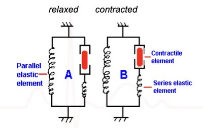
Muscle contractions can be isometric or isotonic, and involve contractile and elastic elements.
Isometric Contraction: Tension without length change.
Isotonic Contraction: Tension with length change.
Concentric: Muscle shortens.
Eccentric: Muscle lengthens.
Mechanical Components:
Contractile Element (CE): Sarcomeres.
Series Elastic Element (SEE): Titin, tendons.
Parallel Elastic Element (PEE): Connective tissue sheaths.
Force Generation: Frequency of Stimulation
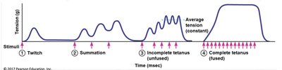
Muscle force depends on the frequency of stimulation, leading to phenomena such as treppe, summation, and tetanus.
Treppe: Stepwise increase in tension with low-frequency stimuli.
Summation: Increased tension with repeated stimulation before relaxation.
Tetanus:
Incomplete: Sustained but shaky contraction.
Complete: Maximal, continuous contraction.
Factors of Force Generation
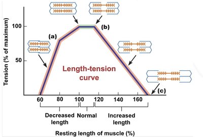
Force generation is influenced by fiber diameter and the length-tension relationship.
Fiber Diameter: Larger fibers generate more force.
Length-Tension Relationship: Optimal sarcomere length allows maximal crossbridge formation.
Optimal: 100-120% resting length.
Excessive stretch: Reduced overlap, less force.
Excessive shortening: Filament interference, less force.
Whole-Muscle Regulation: Recruitment & The Size Principle
Muscle force is regulated by recruiting motor units according to the size principle.
Motor Unit Recruitment: More units recruited for greater force.
Size Principle: Smaller, fatigue-resistant units recruited first; larger units recruited as needed.
Force-Velocity Relationship
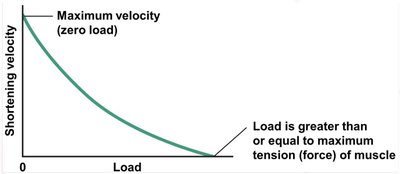
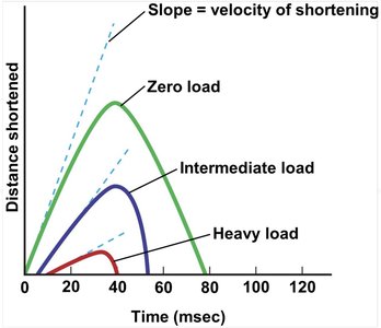
The velocity of muscle shortening is inversely related to the load.
Maximum velocity at zero load.
Velocity decreases as load increases.
Zero velocity at maximum load (isometric).
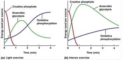
Metabolic Pathways in Skeletal Muscle
Muscle fibers use different metabolic pathways to generate ATP during exercise.
Creatine Phosphate Shuttle: Rapid ATP replenishment for short bursts.
Anaerobic Glycolysis: Sustains high-intensity efforts for 30-60 seconds.
Oxidative Phosphorylation: Supports prolonged, submaximal activity.
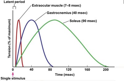
Types of Muscle Fibers
Muscle fibers are classified by contraction speed and metabolic pathway.
Slow Oxidative (Type I): Red, fatigue-resistant, aerobic.
Fast Oxidative-Glycolytic (Type IIA): Pink, intermediate properties.
Fast Glycolytic (Type IIB): White, fast, anaerobic, fatigue-prone.
Most muscles contain a mix of fiber types.
Training Adaptations & Muscular Plasticity
Muscle fibers adapt to training and disuse.
Endurance Training: Increases oxidative capacity; converts fast-glycolytic to fast oxidative-glycolytic.
Resistance Training: Causes hypertrophy (increased size, not number).
Disuse Atrophy: Muscle loss due to inactivity, aging, or disease.
The Cardiovascular System: Cardiac Function
Components of the Cardiovascular System
The cardiovascular system consists of the heart, blood vessels, and blood, working together to transport materials and regulate pressure.
Heart: Muscular pump.
Blood Vessels: Tubular conduits.
Blood: Transport medium.
Heart Anatomy
The heart is protected and anchored by the pericardium and composed of three layers.
Pericardium: Fibrous and serous layers.
Heart Wall: Epicardium, myocardium, endocardium.
Blood Flow through the Heart
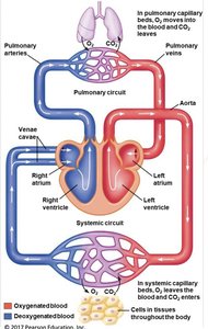
Blood flows through the heart in a specific sequence, ensuring oxygenation and circulation.
Oxygen-poor blood enters the right atrium via vena cava and coronary sinus.
Passes through tricuspid valve to right ventricle.
Through pulmonary valve to pulmonary arteries, then to lungs.
Oxygen-rich blood returns via pulmonary veins to left atrium.
Through mitral valve to left ventricle, then through aortic valve to aorta.
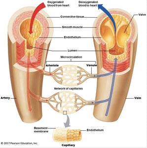
Blood Vessel Anatomy
Blood vessels are specialized for their functions in circulation and exchange.
Arteries: Carry blood away from the heart.
Veins: Return blood to the heart.
Capillaries: Site of exchange; thin walls for permeability.
Microcirculation: Arterioles, capillary network, venules.
Blood Anatomy
Blood is composed of plasma, buffy coat, and erythrocytes.
Plasma: 55% of blood volume.
Buffy coat: Leukocytes and platelets (<1%).
Erythrocytes: 45%; biconcave for flexibility and surface area.
Series vs. Parallel Blood Flow
Blood flow can be arranged in series or parallel, affecting distribution and detoxification.
Series: Sequential flow (e.g., GI tract to liver).
Parallel: Simultaneous flow to multiple organs; allows control and equal distribution.
Electrical Conduction in the Heart
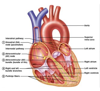
The heart's electrical system coordinates contraction via specialized pathways.
SA node initiates impulse.
Impulse spreads across atria (atrial depolarization).
AV node slows conduction.
AV bundle (bundle of His) transmits impulse.
Bundle branches conduct to ventricles.
Purkinje fibers spread impulse, causing contraction.
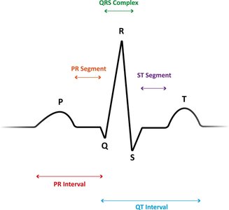
Electrocardiograms (ECG)
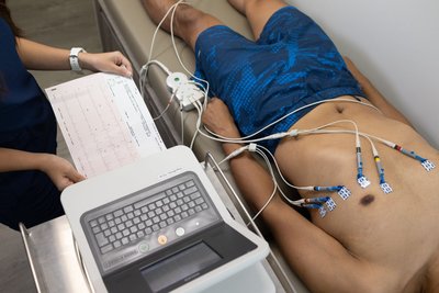
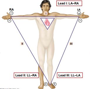
ECGs record the heart's electrical activity and are used to diagnose cardiac conditions.
Leads are placed according to Einthoven's Triangle.
Waveforms:
P-wave: Atrial depolarization.
QRS complex: Ventricular depolarization (hides atrial repolarization).
T-wave: Ventricular repolarization.
The Cardiac Cycle
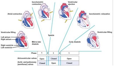
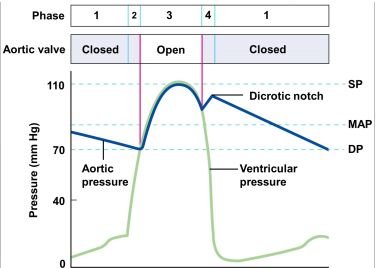
The cardiac cycle consists of systole (contraction) and diastole (relaxation), with changes in pressure and volume.
Atrial pressure peaks between phase 1 and 2.
Ventricular pressure peaks during phase 3.
Aortic pressure drops in phases 1 and 2, peaks in phase 3.
Ventricular Volumes & Ejection Fraction
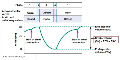
Ventricular volumes and ejection fraction are key measures of cardiac function.
Stroke Volume (SV):
Ejection Fraction (EF):
Cardiac Output
Cardiac output (CO) is the rate at which a ventricle pumps blood.
Formula:
Right and left heart CO must match.
Factors Controlling Cardiac Output
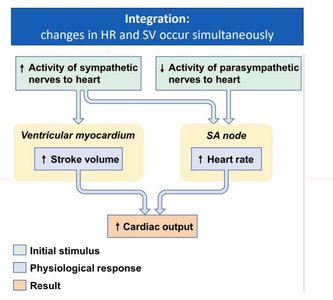
CO is regulated by heart rate (HR) and stroke volume (SV), influenced by intrinsic and extrinsic factors.
Heart Rate: Controlled extrinsically by nervous system and hormones.
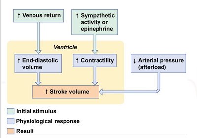
Stroke Volume: Controlled by EDV, contractility, and afterload.
Starling's Law: Increased venous return increases EDV and SV.
Blood Vessels, Blood Flow, and Blood Pressure
Physical Laws of Blood Flow and Pressure
Blood flow is governed by pressure gradients and resistance.
Formula:
Higher pressure difference or lower resistance increases flow.

Bulk Flow
Bulk flow refers to the movement of fluid down a pressure gradient, independent of absolute pressure.
Organ Perfusion Pressure (OPP):
MAP: Mean arterial pressure; CVP: Central venous pressure.
Resistance
Resistance is determined by vessel radius, length, and blood viscosity.
Vessel Radius: Vasoconstriction increases resistance; vasodilation decreases it.
Vessel Length: Longer vessels increase resistance (not significant in humans).
Blood Viscosity: Thicker blood increases resistance.
Total Peripheral Resistance (TPR): Combined resistance of all vessels.
Pulmonary vs. Systemic Flow
Pulmonary and systemic circuits differ in pressure and resistance, but have equal flow.
Pulmonary: Lower pressure and resistance.
Systemic: Higher pressure and resistance.
Blood Vessel Anatomy
Vessels are specialized for their roles in circulation.
Arteries: Thick, elastic, smooth muscle; expand during systole, recoil during diastole.
Arterioles: Smallest arteries, major site of resistance.
Capillaries: Thin, permeable, slow blood flow.
Venules: Smallest veins.
Veins: Thin muscle, valves, large lumen, high compliance.
Pressure in Vessels
Compliance describes how vessels expand in response to pressure.
Arteries: Low compliance, act as pressure reservoir.
Arterioles: Maintain some pressure, large drop-off to protect capillaries.
Measuring Blood Pressure
Blood pressure is measured using a cuff and Korotkoff sounds.
Systolic: Start of turbulent flow.
Diastolic: Return to laminar flow.
Arteriole Tone and Regulation
Arterioles regulate blood flow to capillary beds and MAP via intrinsic and extrinsic mechanisms.
Intrinsic: Local metabolic demand.
Extrinsic: Autonomic nervous system and hormones.
Intrinsic Regulation: Active and Reactive Hyperemia, Myogenic Response
Local mechanisms adjust blood flow based on tissue needs.
Active Hyperemia: Increased metabolic rate triggers vasodilation.
Reactive Hyperemia: Reduced flow triggers vasodilation and overshoot.
Myogenic Response: Stretch triggers constriction; maintains constant flow.
Local Chemical Messengers: Nitric Oxide
Nitric oxide causes vasodilation via cGMP signaling; clinical relevance includes drugs like Viagra.
Extrinsic Control of Arteriolar Radius
Sympathetic and hormonal mechanisms regulate MAP and blood flow distribution.
α receptors: Vasoconstriction in most vessels.
β2 receptors: Vasodilation in liver, heart, skeletal muscle.
Hormonal Control of Arteriolar Radius
Vasopressin (ADH): Vasoconstriction, water reabsorption.
Angiotensin II: Vasoconstriction, stimulates aldosterone, ADH, thirst.
Capillary Anatomy & Exchange
Capillaries are the site of exchange between blood and tissues, with continuous and fenestrated types.
Veins as Volume Reservoir
Veins have high compliance and hold most blood volume at rest; blood shifts to arteries during activity.
Local Control of Capillary Beds
Precapillary sphincters regulate flow into capillary beds, matching local needs.
Venous Return
Venous return is driven by pressure gradients and aided by skeletal muscle and respiratory pumps, blood volume, and venomotor tone.
Regulation of MAP
MAP is regulated by cardiac output and total peripheral resistance.
Formula:
Short-term: Neural (baroreceptor reflex).
Long-term: Hormonal (blood volume).
Neural Control of MAP: Arterial Baroreceptors
Baroreceptors in carotid sinus and aortic arch sense pressure and adjust autonomic output.
Baroreceptor Reflex
Decreased MAP reduces baroreceptor firing, increasing sympathetic and decreasing parasympathetic activity to restore MAP.
Hormonal Control of MAP
Epinephrine: Increases HR, SV, TPR.
Angiotensin II: Vasoconstriction, increases plasma volume.
Vasopressin (ADH): Water conservation, vasoconstriction.
Low Pressure Baroreceptors
Located in large veins and right atrium, these receptors sense blood volume and work with arterial baroreceptors to regulate MAP.
Receptor Type | Location | Senses | Response to Decrease |
|---|---|---|---|
Arterial baroreceptors | Carotid sinus & aortic arch | Arterial pressure (MAP) | ↓ PNS, ↑ SNA |
Low-pressure receptors | Walls of large veins & right atrium | Blood volume | ↑ SNA, ↑ vasopressin secretion |
Additional info: All equations are provided in LaTeX format as required. Images included are only those directly relevant to the adjacent content, reinforcing key concepts in muscle physiology and cardiovascular function.
