 Indietro
Muscle Tissue and Contraction: Study Notes for Anatomy & Physiology
Guida di studio - Note intelligenti
 Appunti personalizzati basati sui tuoi materiali, ampliati con definizioni chiave, esempi e contesto.
Muscle Tissue: Types and Characteristics
Overview of Muscle Tissue
Muscles constitute nearly half of the body’s mass and are essential for converting chemical energy (ATP) into mechanical energy, enabling movement and force generation.
Muscle Terminology: Prefixes such as myo, mys, and sarco refer to muscle.
Three Types of Muscle Tissue:
Skeletal Muscle: Attached to bones, striated, voluntary, contracts rapidly, tires easily, and is powerful.
Cardiac Muscle: Found only in the heart, striated, involuntary.
Smooth Muscle: Located in walls of hollow organs, non-striated, involuntary, visceral.
Muscle Fiber: Only skeletal and cardiac muscles are elongated and referred to as muscle fibers.
Characteristics of Muscle Tissue
Muscle tissue possesses four key properties:
Excitability: Ability to respond to stimuli.
Contractility: Ability to shorten when stimulated.
Extensibility: Ability to be stretched.
Elasticity: Ability to recoil to original length.
Functions of Muscle Tissue
Produce movement (locomotion)
Maintain posture and body position
Stabilize joints
Generate heat
Skeletal Muscle Anatomy
Nerve and Blood Supply
Each skeletal muscle is controlled by nerves (motor units).
Muscles require abundant oxygen and nutrients, and removal of waste products.
Connective Tissue Sheaths
Epimysium: Dense irregular connective tissue surrounding entire muscle.
Perimysium: Fibrous connective tissue surrounding fascicles (groups of muscle fibers).
Endomysium: Fine areolar connective tissue surrounding each muscle fiber.
Muscle Attachments
Muscles attach to bones at two points: origin (immovable) and insertion (movable).
Attachments can be direct (epimysium fused to bone/cartilage) or indirect (via tendons or aponeuroses).
Microscopic Anatomy of Skeletal Muscle
Muscle Fiber Structure
Long, cylindrical cells with multiple nuclei.
Sarcolemma: Plasma membrane of muscle fiber.
Sarcoplasm: Cytoplasm of muscle fiber, contains glycosomes (glycogen storage) and myoglobin (O2 storage).
Myofibrils and Sarcomeres
Myofibrils are densely packed, rodlike elements (~80% of cell volume).
Striations: Alternating dark (A bands) and light (I bands) regions.
Sarcomere: Smallest contractile unit, area between Z discs.
Myofilaments: Actin (thin) and myosin (thick) filaments arranged within sarcomere.
Myofilament Composition
Thick Filaments: Composed of myosin (heavy and light chains), heads form cross bridges during contraction.
Thin Filaments: Composed of actin (G actin and F actin), with regulatory proteins tropomyosin and troponin.
Other structural proteins: Titin (elastic filament), Dystrophin, Nebulin, Myomesin, C proteins.
Calcium Regulation and Muscle Contraction
Sarcoplasmic Reticulum (SR) and T Tubules
SR: Network of smooth ER surrounding myofibrils, regulates intracellular Ca2+ levels.
T Tubules: Extensions of sarcolemma, allow electrical signals to penetrate deep into muscle fiber.
Triad: Area formed by one T tubule and two terminal cisterns of SR.
Sliding Filament Model of Contraction
During contraction, thin filaments slide past thick filaments, increasing overlap.
Myosin heads bind to actin, forming cross bridges, and pull actin toward the center of the sarcomere.
Muscle fiber shortens as Z discs are pulled toward M line, I bands shorten, H zones disappear, and A bands move closer.
Muscle Fiber Contraction: Steps and Mechanisms
Neuromuscular Junction and Action Potential
Motor neurons stimulate muscle fibers via the neuromuscular junction (NMJ).
NMJ consists of axon terminals, synaptic cleft, and junctional folds with ACh receptors.
Action potential (AP) crosses from neuron to muscle cell via acetylcholine (ACh).
Ion Channels
Chemically gated ion channels: Opened by neurotransmitters (e.g., ACh).
Voltage-gated ion channels: Open or close in response to changes in membrane potential.
Four Steps of Muscle Contraction
Events at Neuromuscular Junction: AP arrives, Ca2+ enters neuron, ACh released, binds to receptors, Na+ enters muscle fiber, end plate potential generated.
Muscle Fiber Excitation: Sarcolemma depolarizes, AP propagates.
Excitation-Contraction Coupling: AP travels down T tubules, Ca2+ released from SR, contraction initiated.
Cross Bridge Cycling: Ca2+ binds to troponin, tropomyosin moves, myosin binds to actin, ATP powers cycle.
Rigor Mortis
After death, ATP production ceases, Ca2+ accumulates, cross bridges form and cannot detach, resulting in muscle stiffness.
Muscle Contraction: Types and Responses
Isometric vs. Isotonic Contractions
Isometric: Muscle tension increases, but muscle does not shorten.
Isotonic: Muscle shortens and moves load; includes concentric (shortening) and eccentric (lengthening) contractions.
Motor Units and Muscle Twitch
Motor Unit: Motor neuron and all muscle fibers it innervates.
Muscle Twitch: Response to a single AP; consists of latent, contraction, and relaxation phases.
Graded Muscle Responses
Strength of contraction varies by frequency and intensity of stimulation.
Wave Summation: Rapid stimuli increase force.
Tetanus: Sustained contraction (unfused or fused).
Recruitment: More motor units activated for stronger contractions.
Energy for Muscle Contraction
ATP Regeneration Mechanisms
Direct Phosphorylation: Creatine phosphate donates phosphate to ADP to form ATP.
Anaerobic Pathway: Glycolysis and lactic acid formation (no oxygen required).
Aerobic Pathway: Occurs in mitochondria, requires oxygen, produces CO2, H2O, and up to 32 ATP per glucose.
Energy Systems During Exercise
Aerobic Endurance: Duration muscle contracts using aerobic pathways.
Anaerobic Threshold: Point where metabolism shifts to anaerobic pathway.
Muscle Fatigue and Recovery
Causes and Recovery
Fatigue: Inability to contract despite stimulation; caused by ionic imbalances, increased inorganic phosphate, or impaired calcium release.
Recovery: Oxygen reserves replenished, lactic acid reconverted, glycogen restored, ATP and creatine phosphate resynthesized.
EPOC: Excess postexercise oxygen consumption (formerly "oxygen debt").
Force, Speed, and Adaptation of Muscle Contraction
Factors Affecting Force
Number of fibers stimulated (recruitment)
Relative size of fibers (hypertrophy)
Frequency of stimulation
Degree of muscle stretch (optimal sarcomere length: 80–120% resting length)
Adaptation to Exercise
Aerobic Exercise: Increases capillaries, mitochondria, myoglobin; improves endurance and resistance to fatigue.
Resistance Exercise: Increases fiber size (hypertrophy), mitochondria, myofilaments, glycogen, connective tissue; improves strength and size.
Muscle Tone and Atrophy
Muscle tone: Constant, slight contraction due to spinal reflexes.
Disuse atrophy: Loss of mass due to immobilization or neural loss; can be rapid and irreversible.
Smooth Muscle
Structure and Function
Found in walls of hollow organs (except heart).
Organized into sheets; two layers (longitudinal and circular) for mixing and propulsion.
Spindle-shaped, single nucleus, no striations.
Contains only endomysium; lacks other connective tissue sheaths.
Innervated by autonomic nervous system; neurotransmitters released via varicosities.
Electrically connected via gap junctions.
Contains thick and thin filaments, but no sarcomeres; filaments arranged diagonally for corkscrew contraction.
Dense bodies anchor filaments; correspond to Z discs in skeletal muscle.
No troponin; calmodulin binds Ca2+.
Muscle Development and Disease
Sex Differences and Muscular Dystrophy
Male skeletal muscle: 42% body mass; female: 36% (due to testosterone).
Muscular dystrophy: Group of inherited diseases causing muscle degeneration.
Duchenne Muscular Dystrophy (DMD): Most common, sex-linked, lack of dystrophin, leads to muscle weakness and early death.
Study Aids and Interactive Resources
Using Online Study Tools
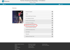
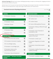
Students are encouraged to use interactive physiology resources and study areas for additional practice and understanding.

Summary Table: Types of Muscle Tissue
Type | Location | Striations | Control | Key Features |
|---|---|---|---|---|
Skeletal | Bones | Yes | Voluntary | Rapid, powerful, tires easily |
Cardiac | Heart | Yes | Involuntary | Rhythmic contractions |
Smooth | Hollow organs | No | Involuntary | Slow, sustained contractions |
Summary Table: ATP Regeneration Pathways
Pathway | Location | Oxygen Required? | ATP Yield | Duration |
|---|---|---|---|---|
Direct Phosphorylation | Cytoplasm | No | 1 ATP per CP | 15 seconds |
Anaerobic Glycolysis | Cytoplasm | No | 2 ATP per glucose | 30–40 seconds |
Aerobic Respiration | Mitochondria | Yes | 32 ATP per glucose | Hours |
Summary Table: Steps of Muscle Contraction
Step | Description |
|---|---|
1. Neuromuscular Junction | AP arrives, ACh released, end plate potential generated |
2. Muscle Fiber Excitation | Depolarization and AP propagation |
3. Excitation-Contraction Coupling | AP triggers Ca2+ release, contraction initiated |
4. Cross Bridge Cycling | Myosin binds actin, ATP powers cycle |
