 Indietro
Muscle Tissue and Physiology: Structure, Function, and Types
Guida di studio - Note intelligenti
 Appunti personalizzati basati sui tuoi materiali, ampliati con definizioni chiave, esempi e contesto.
Muscle Tissue and Physiology
Overview of Muscle Tissue
Muscle tissue is essential for generating force, movement, and maintaining various physiological functions. There are three primary types of muscle tissue: skeletal, cardiac, and smooth muscle. Each type has unique structural and functional characteristics.
Generating force (muscle tension): All muscle tissues produce tension, which is fundamental for movement and stability.
Other functions: Movement, posture maintenance, joint stabilization, heat generation, and regulation of material flow through hollow organs.
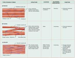
Types of Muscle Tissue
The three types of muscle tissue differ in structure, location, control, and function. The following table summarizes these differences:
Type | Structure | Location | Voluntary/Involuntary | Function |
|---|---|---|---|---|
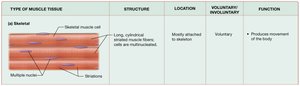
Skeletal | Long, cylindrical, striated fibers; multinucleated | Attached to skeleton | Voluntary | Produces movement of the body |
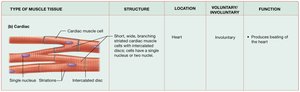
Cardiac | Short, branching, striated fibers; single nucleus; intercalated discs | Heart | Involuntary | Produces beating of the heart |
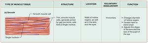
Smooth | Thin, smooth fibers; single nucleus; no striations | Walls of hollow organs, skin, eyes | Involuntary | Changes diameter of tubes, causes hair to stand erect, adjusts shape of lens and pupil |
Properties of Muscle Cells
Muscle cells possess specialized properties that enable their function:
Contractility: Ability to contract; proteins in the cell draw closer together, not always resulting in cell shortening.
Excitability: Ability to respond to stimuli (chemical, mechanical, or electrical).
Conductivity: Ability to conduct electrical changes across the plasma membrane.
Extensibility: Ability to stretch up to three times resting length without rupturing.
Elasticity: Ability to return to original length after stretching.
Structure of Muscle Cells
Muscle Cell Terminology and Organization
Muscle cells, also known as myocytes, have specialized structures and terminology:
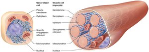
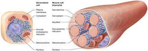
Sarcoplasm: Cytoplasm of the muscle cell.
Sarcolemma: Plasma membrane of the muscle cell.
Sarcoplasmic reticulum (SR): Modified endoplasmic reticulum forming a web-like network around myofibrils; stores and releases calcium ions.
Myofibrils: Bundles of contractile proteins; most abundant organelle in muscle cells.
Myofibril Arrangement
Myofibrils are composed of myofilaments, which are made up of contractile, regulatory, and structural proteins. The arrangement of myofibrils differs between muscle types, with smooth muscle having a unique organization compared to skeletal and cardiac muscle.
Structure and Function of Skeletal Muscle Fibers
Skeletal Muscle Fiber Characteristics
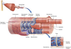
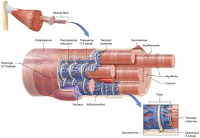
Skeletal muscle fibers are long, cylindrical cells surrounded by endomysium. They are formed by the fusion of embryonic myoblasts, resulting in multinucleated cells. Myofibrils are the most abundant organelle, and the sarcoplasmic reticulum surrounds them, storing and releasing calcium ions.
Length: Up to 30 cm
Thickness: Up to 100 μm

Internal Structure of Skeletal Muscle Fiber
Key internal structures include:
Transverse tubules (T-tubules): Deep extensions of the sarcolemma that surround each myofibril and form a tunnel-like network filled with extracellular fluid.
Terminal cisternae: Enlarged sections of the SR that flank each T-tubule; two terminal cisternae plus a T-tubule form a triad.
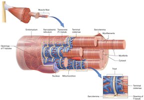
Structure of the Myofibril
Myofilaments and Their Proteins
Myofibrils are composed of hundreds to thousands of myofilaments, which include:
Contractile proteins: Generate tension (e.g., actin and myosin).
Regulatory proteins: Dictate when a fiber may contract (e.g., troponin and tropomyosin).
Structural proteins: Maintain proper alignment and stability.
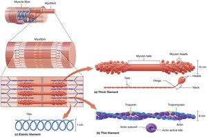
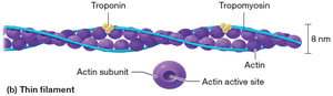
Thin Filaments: Tropomyosin and Troponin
Thin filaments are primarily composed of actin, with regulatory proteins:
Tropomyosin: Long, rope-like protein that twists around actin, covering active sites.
Troponin: Small, globular protein that holds tropomyosin in place and assists in turning contractions on and off.
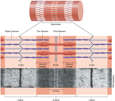
Myofilament Arrangement and the Sarcomere
Sarcomere Structure and Bands
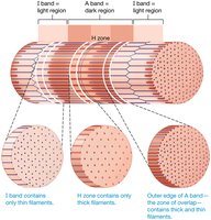
The sarcomere is the functional unit of muscle contraction, defined by alternating light and dark bands:
I band: Contains only thin filaments.
Z disc: Structural proteins anchoring thin filaments and serving as attachment points for elastic filaments.
A band: Contains both thick and thin filaments; generates tension during contraction.
H zone: Middle of A band with only thick filaments.
M line: Structural proteins holding thick filaments in place and anchoring elastic filaments.
Levels of Organization within a Skeletal Muscle
Hierarchical Structure
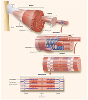
Skeletal muscle is organized in a hierarchical manner:
Muscle → Fascicle → Muscle fiber → Myofibril → Myofilament
Each level is surrounded by connective tissue and contributes to overall muscle function.
Duchenne Muscular Dystrophy (DMD)
Overview and Effects
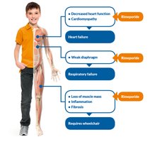
DMD is a genetic disorder characterized by progressive muscle degeneration and weakness. It affects skeletal, cardiac, and respiratory muscles, leading to severe complications and loss of mobility.
Symptoms: Decreased heart function, cardiomyopathy, heart failure, weak diaphragm, respiratory failure, loss of muscle mass, inflammation, fibrosis, and wheelchair dependence.
The Sliding-Filament Mechanism of Contraction
Mechanism of Muscle Contraction
The sliding-filament mechanism explains how muscle contraction occurs:
Sarcomere: Extends from one Z-disc to the next; functional unit of contraction.
Contraction: Myosin heads attach to actin, pulling thin filaments toward the M line, shortening the sarcomere.
Band changes: I band and H zone narrow, A band remains unchanged.
Whole muscle contraction: Sarcomeres contract simultaneously, shortening the entire muscle fiber.
*Additional info: The sliding-filament mechanism is fundamental to understanding muscle physiology and is a key concept in muscle contraction.*
