 Indietro
Muscle Tissue and Physiology: Structured Study Notes
Guida di studio - Note intelligenti
 Appunti personalizzati basati sui tuoi materiali, ampliati con definizioni chiave, esempi e contesto.
Muscle Tissue
Three Types of Muscle Tissue
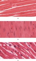
Muscle tissue is classified into three main types, each with distinct structural and functional characteristics:
Skeletal muscle: Voluntary control, multinucleated, striated appearance.
Smooth muscle: Involuntary control, uninucleated, non-striated.
Cardiac muscle: Involuntary control, uninucleated, striated, found only in the heart.
Special Characteristics of Muscle Tissue
Muscle tissue possesses unique properties that enable its function:
Excitability (responsiveness): Ability to receive and respond to stimuli.
Contractility: Ability to shorten when stimulated.
Extensibility: Ability to be stretched.
Elasticity: Ability to recoil to resting length after stretching.
Muscle Functions
Muscle tissue serves several essential functions in the body:
Movement: Facilitates movement of bones or fluids (e.g., blood).
Posture: Maintains posture and body position.
Joint stabilization: Stabilizes joints.
Heat generation: Especially by skeletal muscle, helps maintain body temperature.
Skeletal Muscle Structure
Connective Tissue Sheaths
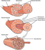
Skeletal muscle is organized and protected by three layers of connective tissue:
Epimysium: Surrounds the entire muscle.
Perimysium: Surrounds fascicles (bundles of muscle fibers).
Endomysium: Surrounds individual muscle fibers.
Muscle Attachments
Muscles attach to bones or cartilage in two ways:
Direct attachment: Epimysium fuses with periosteum or perichondrium.
Indirect attachment: Connective tissue extends as a tendon or aponeurosis.
Microscopic Anatomy of Skeletal Muscle Fiber
Muscle Fiber Structure
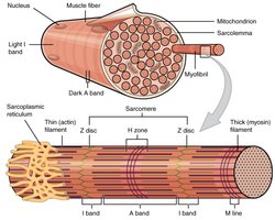
Skeletal muscle fibers are large, multinucleated cells with specialized organelles:
Multiple peripheral nuclei
Many mitochondria
Glycosomes: Glycogen storage
Myoglobin: Oxygen storage
Myofibrils, sarcoplasmic reticulum, and T tubules
Sarcomere: The Functional Unit
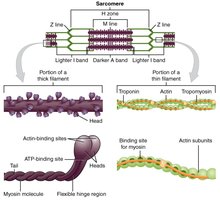
The sarcomere is the smallest contractile unit of muscle fiber, defined as the region between two Z discs. It is composed of thick (myosin) and thin (actin) filaments.
Features of a Sarcomere
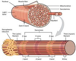
Thick (Myosin) filaments: Run the length of the A band; have heads with binding sites for actin and ATP.
Thin (Actin) filaments: Run the length of the I band and partway into the A band; contain tropomyosin and troponin (regulatory proteins).
Z disc: Anchors thin filaments and connects myofibrils.
H zone: Lighter mid-region where filaments do not overlap.
M line: Holds adjacent thick filaments together.
T Tubules and Sarcoplasmic Reticulum
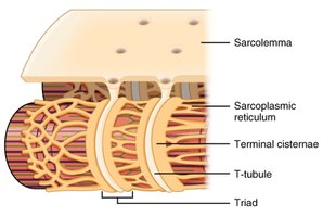
T tubules are extensions of the sarcolemma that penetrate the cell's interior at each A band-I band junction. The sarcoplasmic reticulum is a network of smooth endoplasmic reticulum surrounding each myofibril, regulating intracellular Ca2+ levels.
Muscle Contraction
Sliding Filament Model
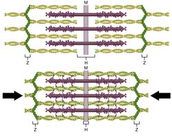
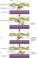
Muscle contraction is based on the sliding filament model, where myosin heads bind to actin, detach, and rebind, propelling thin filaments toward the M line. This shortens the sarcomere, muscle cell, and whole muscle.
Requirements for Skeletal Muscle Contraction
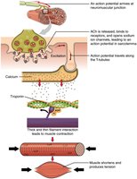
Activation: Neural stimulation at the neuromuscular junction.
Excitation-contraction coupling: Generation and propagation of an action potential along the sarcolemma.
Final trigger: Brief rise in intracellular Ca2+ levels.
Events at the Neuromuscular Junction
Skeletal muscles are stimulated by somatic motor neurons. Each axon forms several branches as it enters a muscle, and each axon ending forms a neuromuscular junction with a single muscle fiber.
Power Stroke Sequence
The power stroke is the process by which myosin heads pull actin filaments, using ATP for energy. This cycle repeats during muscle contraction.
Types of Muscle Contractions
Isometric vs. Isotonic Contractions
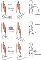
Isometric: Muscle tension increases but does not exceed the load; no change in muscle length.
Isotonic: Muscle length changes when load is placed on it.
Concentric: Muscle shortens and does work.
Eccentric: Muscle contracts as it lengthens.
Motor Unit and Muscle Twitch
Motor Unit
A motor unit consists of one motor neuron and all the muscle fibers it supplies. Small motor units control fine movements, while large motor units control weight-bearing muscles. Motor units contract asynchronously to prevent fatigue.
Muscle Twitch
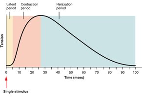
A muscle twitch is the response of a muscle to a single, brief threshold stimulus. It consists of three phases:
Latent period: Events of excitation-contraction coupling.
Period of contraction: Cross bridge formation; tension increases.
Period of relaxation: Ca2+ reentry into the SR; tension declines to zero.
Graded Muscle Responses
Frequency and Strength of Stimulation
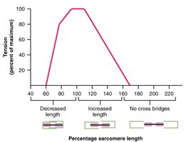
Muscle contraction strength is graded by changing the frequency and strength of stimulation. Maximal tension occurs at 80-120% of a sarcomere's resting length.
Wave Summation and Tetanus
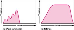
Wave summation: Fibers are stimulated while previous twitch is still occurring, increasing tension.
Tetanus: Continuous contraction; all sarcomeres form cross-bridges.
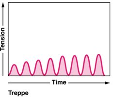
Treppe
Treppe is the phenomenon where muscle tension increases in a graded manner after periods of dormancy, as contractions become more efficient with steady ATP supply.
Response to Change in Stimulus Strength
Recruitment: The process of increasing the number of active motor units to control contraction force.
Threshold stimulus: The minimum stimulus required for contraction.
Maximum stimulation: The strongest stimulus that recruits all motor units.
Muscle Metabolism: Energy for Contraction
ATP Regeneration
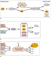
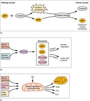
ATP is the only direct energy source for muscle contraction. It is regenerated by three methods:
Direct phosphorylation of ADP by creatine phosphate (CP)
Anaerobic pathway (glycolysis)
Aerobic respiration
Muscle Metabolism Details
Creatine phosphate: Stores energy in phosphate bonds; provides energy for about 15 seconds.
Glycolysis: Anaerobic process; slower rate of ATP availability.
Aerobic respiration: Provides 95% of ATP for resting or moderately active muscles.
Oxygen debt: The amount of oxygen needed to restore ATP and creatine phosphate levels and convert lactic acid to pyruvic acid or glucose/glycogen in the liver.
Muscle Fatigue
Muscle fatigue is the physiological inability to contract, occurring when ATP production fails to keep pace with use, ionic imbalances are present, or oxygen debt results.
Classes of Muscle Fibers
Types of Muscle Fibers
Type | Characteristics | Function |
|---|---|---|
Type I | Abundant mitochondria, myoglobin, capillaries; aerobic respiration | Endurance activities (e.g., marathons) |
Type II | Low mitochondria, myoglobin, capillaries; vast sarcoplasmic reticulum | Anaerobic respiration; fatigue quickly |
Type IIA | Fast oxidative (pink); more resistant to fatigue | Sustained power activities (e.g., basketball, soccer) |
Type IIB | Fast twitch (white); recruited for short-duration, high-intensity bursts | Bursts of power (e.g., maximal lifts, sprints) |
Quiz:
Smooth muscle tissues have varicosities
Cardiac muscles have intercalated discs
Treppe muscle contraction helps someone with a "warm up" before a workout
Holding a plank for 30 seconds would be considered a isometric contraction
The 'key' and the 'key hole' to muscle contraction is made up of calcium; troponin
Triad is made up of two terminal cisternae and the T-Tubule
The neuromuscular junction involves the neurotransmitter, acetylcholine
Additional info: Muscle fiber types are recruited based on activity intensity and duration, with Type I fibers used for endurance, and Type II fibers for rapid, powerful movements.
