 Indietro
Muscle Tissue and Skeletal Muscle Structure: Study Notes
Guida di studio - Note intelligenti
 Appunti personalizzati basati sui tuoi materiali, ampliati con definizioni chiave, esempi e contesto.
Muscle Tissue
Overview of Muscle Tissue Types
Muscle tissue is a primary tissue type in the human body, essential for movement and force generation. It is divided into three main types:
Skeletal muscle: Voluntary, striated muscle attached to bones.
Cardiac muscle: Involuntary, striated muscle found in the heart.
Smooth muscle: Involuntary, non-striated muscle found in walls of hollow organs.
All muscle tissues share the following properties:
Excitability: Ability to respond to stimuli.
Elasticity: Ability to recoil to original length after stretching.
Contractility: Ability to shorten forcefully.
Skeletal Muscle
Functions of Skeletal Muscle
Produce skeletal movement
Maintain posture and body position
Support soft tissues
Guard entrances and exits (e.g., sphincters)
Maintain body temperature (through heat production)
Store nutrient reserves (e.g., glycogen)
Organization of Skeletal Muscle
Epimysium: Outer layer surrounding the entire muscle.
Perimysium: Surrounds bundles of muscle fibers (fascicles).
Endomysium: Surrounds individual muscle fibers.
Tendons: Connect muscle to bone; aponeuroses are broad, flat tendons.
Skeletal Muscle Fibers
Very long, cylindrical, multinucleated cells.
Develop from fusion of myoblasts (mesodermal cells).
Striated and under voluntary control.
Key Characteristics of Muscle Fibers
Sarcolemma: Excitable plasma membrane of muscle fiber.
T-tubules: Invaginations of sarcolemma that transmit action potentials.
Sarcoplasmic reticulum (SR): Stores calcium ions (Ca2+); terminal cisternae are enlarged areas of SR.
Triad: One T-tubule flanked by two terminal cisternae.
Sarcoplasm: Cytoplasm of muscle fiber.
Myofibrils: Bundles of protein filaments (myofilaments) responsible for contraction.
Structure of the Sarcomere
Sarcomere Organization
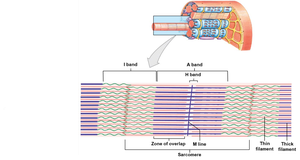
The sarcomere is the basic contractile unit of muscle fiber, composed of repeating patterns of thick and thin filaments.
A Band: Dark band; contains thick filaments and overlapping thin filaments.
M Line: Center of the A band; midline of sarcomere.
H Band: Area around the M line; contains thick filaments only.
Zone of overlap: Region where thick and thin filaments overlap.
I Band: Light band; contains only thin filaments.
Z Lines: Boundaries between adjacent sarcomeres.
Titin: Elastic protein that stabilizes filaments and prevents overstretching.
Myofilaments
Thin filaments: Primarily composed of actin, with regulatory proteins:
F-actin: Two twisted rows of globular G-actin.
Nebulin: Holds F-actin strands together.
Tropomyosin: Blocks actin-myosin interaction at rest.
Troponin: Binds tropomyosin to G-actin; regulated by Ca2+.
Thick filaments: Composed of myosin molecules.
Myosin tail: Binds to other myosin molecules.
Myosin head: Interacts with actin to form cross-bridges.
Muscle Contraction
Sliding Filament Theory
Muscle contraction occurs as thin filaments slide past thick filaments, shortening the sarcomere without changing the length of the filaments themselves.
Thin filaments slide toward the M line.
The width of the A band remains constant.
Z lines move closer together.
Neuromuscular Junction
Presynaptic terminal: Axon of motor neuron containing synaptic vesicles with acetylcholine (ACh).
Synaptic cleft: Space between neuron and muscle fiber; contains acetylcholinesterase enzyme.
Postsynaptic membrane: Motor end-plate with ligand-gated Na+ channels that open in response to ACh.
Excitation-Contraction Coupling
Action potential travels down T-tubules to triads.
Opens voltage-gated Ca2+ channels on SR, releasing Ca2+ into sarcoplasm.
Ca2+ triggers contraction by binding to troponin.
Cross-Bridge Cycle
Ca2+ binds to troponin, causing tropomyosin to move and expose active sites on actin.
Myosin heads form cross-bridges with actin.
Powerstroke: Myosin head pivots, pulling actin filament.
Cross-bridge detachment: ATP binds to myosin, causing it to release actin.
Reactivation: Myosin head hydrolyzes ATP and returns to cocked position.
Skeletal Muscle Relaxation
Ca2+ is pumped back into SR.
Ca2+ detaches from troponin; tropomyosin covers active sites.
Muscle returns to resting state.
Rigor mortis: Stiffening of muscles after death due to lack of ATP.
Tension and Types of Contraction
Phases of a Muscle Twitch
Latent period: Action potential moves through sarcolemma; Ca2+ released.
Contraction phase: Ca2+ binds; tension builds to peak.
Relaxation phase: Ca2+ levels fall; tension returns to resting.
Summation and Tetanus
Wave summation: Increased tension from repeated stimulation before relaxation completes.
Complete tetanus: High-frequency stimulation prevents relaxation, causing continuous contraction.
Motor Units and Tension Production
Each motor unit contains multiple muscle fibers controlled by a single neuron.
Recruitment: Increasing the number of active motor units increases tension.
Maximum tension: All motor units in tetanus; can only be sustained briefly.
Sustained tension: Less than maximum; allows rotation and rest of motor units.
Muscle tone: Baseline tension in resting muscle; maintains posture and readiness.
Types of Contractions
Isotonic contraction: Muscle changes length, causing movement.
Concentric: Muscle tension > load; muscle shortens.
Eccentric: Muscle tension < load; muscle lengthens.
Isometric contraction: Muscle develops tension but does not change length.
Muscle Relaxation and Return to Resting Length
Elastic forces: Tendons and ligaments help restore sarcomere length.
Opposing muscle contractions: Antagonistic muscles reverse movement.
Energy for Muscle Contraction
ATP and Creatine Phosphate (CP) Reserves
ATP: Immediate energy source for contraction.
Creatine phosphate (CP): Stores excess energy to quickly regenerate ATP.
Enzyme creatine kinase (CK) catalyzes transfer of phosphate from CP to ADP to form ATP.
ATP Generation Pathways
Aerobic metabolism: Occurs in mitochondria; uses fatty acids and glucose; produces up to 34 ATP per glucose.
Anaerobic glycolysis: Occurs in cytoplasm; breaks down glucose from glycogen; produces 2 ATP per glucose and lactic acid as a by-product.
Energy Use and Muscle Activity
At rest: Muscles metabolize fatty acids and store glycogen.
At peak activity: Anaerobic glycolysis predominates, leading to lactic acid buildup.
Muscle Fatigue
Occurs when muscles can no longer perform required activity.
Causes: Depletion of metabolic reserves, damage to sarcolemma/SR, low pH (lactic acid), muscle exhaustion, and pain.
