 Indietro
Muscle Tissue: Structure and Contraction Mechanisms
Guida di studio - Note intelligenti
 Appunti personalizzati basati sui tuoi materiali, ampliati con definizioni chiave, esempi e contesto.
Muscle Tissue: Structure and Contraction Mechanisms
Structure of Skeletal Muscle Cells
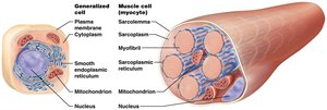
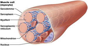
Skeletal muscle cells, also known as myocytes or muscle fibers, possess specialized structures that enable contraction and force generation. These cells are elongated and multinucleated, containing unique organelles and membranes adapted for their function.
Sarcoplasm: The cytoplasm of a muscle cell, containing dissolved proteins, ions, and organelles.
Sarcolemma: The plasma membrane of the muscle cell, crucial for conducting electrical signals.
Sarcoplasmic Reticulum (SR): A modified smooth endoplasmic reticulum forming a network around myofibrils, responsible for storing and releasing calcium ions.
Myofibrils: Cylindrical bundles of contractile proteins, making up 50–80% of cell volume and enabling contraction.
Mitochondria: Provide ATP for muscle contraction.
Nucleus: Multiple nuclei are present due to the cell's large size and high metabolic demand.
Transverse Tubules and Triads
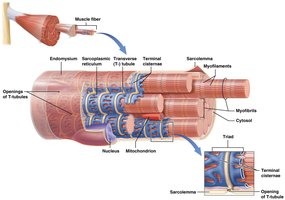
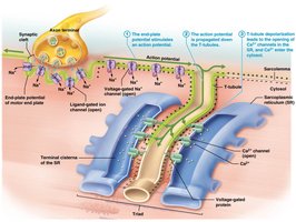
Transverse (T) tubules are deep invaginations of the sarcolemma that surround each myofibril, forming a tunnel-like network filled with extracellular fluid. Terminal cisternae are enlarged regions of the SR flanking each T-tubule. Together, two terminal cisternae and a T-tubule form a triad, which is essential for rapid transmission of action potentials and release of calcium ions.
Organization of Myofibrils and Myofilaments
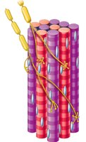
Each myofibril consists of hundreds to thousands of myofilaments, which are classified into three types:
Thick filaments: Composed of the contractile protein myosin.
Thin filaments: Composed of contractile and regulatory proteins actin, tropomyosin, and troponin.
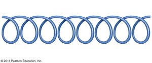
Elastic filaments: Composed of the structural protein titin, which stabilizes the myofibril and resists excessive stretching.
Contractile, Regulatory, and Structural Proteins
Contractile proteins: Myosin (thick filaments) and actin (thin filaments) produce tension.
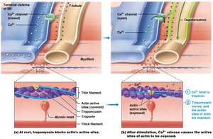
Regulatory proteins: Tropomyosin and troponin control when contraction occurs by covering or exposing actin's active sites.
Structural proteins: Titin maintains stability and alignment of myofilaments.
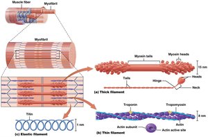
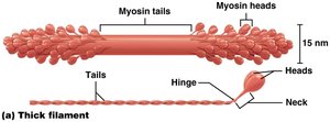
Thick Filaments (Myosin)
Thick filaments are made of myosin molecules, each with a globular head, tail, hinge, and neck. The myosin head contains an active site for binding actin and ATP hydrolysis.
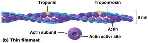
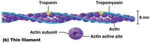
Thin Filaments (Actin, Tropomyosin, Troponin)
Thin filaments consist of actin subunits forming two intertwined strands. Each actin has an active site for myosin binding. Tropomyosin is a rope-like protein that covers actin's active sites, while troponin binds to actin, tropomyosin, and Ca2+, regulating contraction.
Elastic Filaments (Titin)
Elastic filaments are composed of titin, a spring-like protein that stabilizes myofibril structure and resists overstretching. Titin runs through the core of the thick filament, uncoiling when stretched and recoiling when the force is removed.
Myofilament Arrangement and the Sarcomere
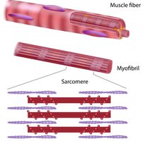
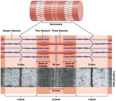
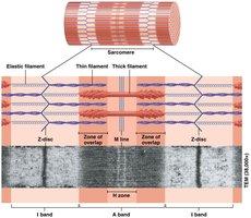
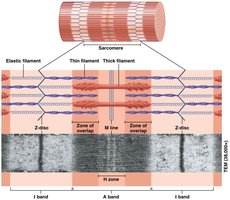
The arrangement of myofilaments creates the striated appearance of skeletal muscle. The sarcomere is the functional unit of contraction, defined by the region between two Z-discs.
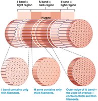
I band: Contains only thin filaments.
A band: Contains both thick and thin filaments, including the zone of overlap.
H zone: Contains only thick filaments, located in the middle of the A band.
Z disc: Anchors thin filaments and titin, and connects adjacent sarcomeres.
M line: Anchors thick filaments and titin in the center of the sarcomere.
Sliding Filament Theory of Muscle Contraction
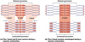
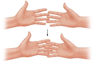
Muscle contraction occurs through the sliding filament mechanism. Myosin heads bind to actin and pull the thin filaments toward the M line, shortening the sarcomere. The I band and H zone narrow, while the A band remains unchanged. This process does not shorten the filaments themselves, but rather their relative position changes.
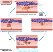
Power stroke: Myosin head pivots, pulling actin toward the center.
Crossbridge cycle: Repeated attachment, pivot, and detachment of myosin heads as long as ATP and Ca2+ are available.
Anatomy of the Neuromuscular Junction (NMJ)
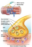
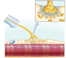
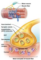
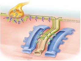
The neuromuscular junction is the synapse where a motor neuron communicates with a muscle fiber. It transmits the nerve impulse (action potential) to the sarcolemma, initiating contraction.
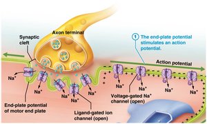
Axon terminal: Contains synaptic vesicles filled with acetylcholine (ACh).
Synaptic cleft: Space between axon terminal and muscle fiber, filled with collagen and gel.
Motor end plate: Specialized region of sarcolemma with ligand-gated Na+ channels.
Physiology of Skeletal Muscle Contraction
Muscle contraction is divided into three phases: excitation, excitation-contraction coupling, and contraction (crossbridge cycle).
Excitation phase: Action potential arrives at NMJ, ACh is released, and Na+ enters the muscle cell.
Excitation-contraction coupling: Action potential travels along sarcolemma and T-tubules, triggering Ca2+ release from SR.
Contraction phase: Ca2+ binds to troponin, tropomyosin moves, exposing actin's active sites for myosin binding.
Membrane Potential and Action Potentials
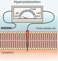
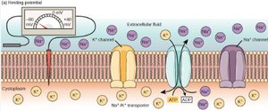
The resting membrane potential of a muscle fiber is -85 mV. Action potentials are generated by opening ligand-gated and voltage-gated ion channels, resulting in brief changes from negative to positive and back.
Depolarization: Na+ enters, making the membrane more positive.
Repolarization: K+ exits, restoring negative potential.
Excitation-Contraction Coupling
When an action potential reaches the triad, Ca2+ is released from the terminal cisternae of the SR. Ca2+ binds to troponin, causing tropomyosin to move and expose actin's active sites, allowing contraction to begin.
Summary Table: Sarcomere Regions and Filament Types
Region | Filament Type | Function |
|---|---|---|
I band | Thin filaments | Light region, narrows during contraction |
A band | Thick & thin filaments | Dark region, remains unchanged |
H zone | Thick filaments | Middle of A band, narrows during contraction |
Z disc | Structural proteins | Anchors thin filaments, titin, and connects sarcomeres |
M line | Structural proteins | Anchors thick filaments and titin |
Key Equations
Resting Membrane Potential:
ATP Hydrolysis (for crossbridge cycle):
Additional info:
These notes expand on the original content by providing definitions, examples, and context for each structure and process, ensuring completeness and academic quality for exam preparation.
