 Indietro
Muscle Tissue: Structure and Function in Anatomy & Physiology
Guida di studio - Note intelligenti
 Appunti personalizzati basati sui tuoi materiali, ampliati con definizioni chiave, esempi e contesto.
Muscle Tissue Overview
Types of Muscle Tissue
Muscle tissue is essential for movement, stability, and various physiological functions. There are three primary types of muscle tissue, each with distinct characteristics and roles:
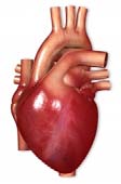
Cardiac Muscle: Striated and involuntary, found only in the heart. Responsible for pumping blood throughout the body.
Smooth Muscle: Non-striated and involuntary, located in the walls of internal organs such as the digestive tract and blood vessels.
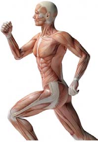
Skeletal Muscle: Striated and voluntary, attached to bones and responsible for body movement, posture, and heat production.
Functions of Skeletal Muscle
Produce skeletal movement: Muscles contract to move bones.
Maintain body position: Continuous muscle contractions stabilize joints and posture.
Support soft tissues: Muscles shield internal organs.
Guard openings: Muscles control entrances and exits of the body (e.g., sphincters).
Maintain body temperature: Muscle activity generates heat.
Store nutrient reserves: Muscles store glycogen and proteins for energy.
Connective Tissue Layers in Skeletal Muscle
Three Layers of Connective Tissue
Skeletal muscle is organized into three connective tissue layers that provide structure, support, and facilitate repair:
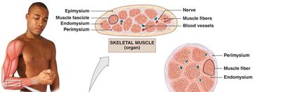
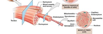
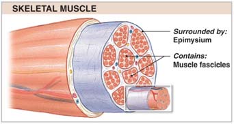
Epimysium: The outermost layer, composed of collagen, surrounds the entire muscle and separates it from surrounding tissues.
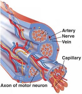
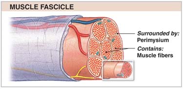
Perimysium: Surrounds bundles of muscle fibers called fascicles, contains blood vessels and nerves.
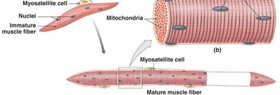
Endomysium: Encloses individual muscle fibers, contains capillaries, nerve fibers, and myosatellite cells (muscle stem cells).
Muscle Attachments
The connective tissue layers converge at the ends of muscles to form attachments:
Tendon: A bundle of connective tissue attaching muscle to bone.
Aponeurosis: A sheet-like connective tissue attachment.
Nerves and Blood Vessels
Skeletal muscles are highly vascularized and innervated:
Nerves: Control voluntary muscle contraction via signals from the central nervous system (CNS).
Blood Vessels: Supply oxygen and nutrients, remove metabolic waste.
Structure of Skeletal Muscle Fibers
Development and Organization
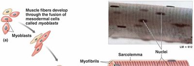
Skeletal muscle fibers are formed by the fusion of myoblasts and contain multiple nuclei. Mature fibers are long, cylindrical cells packed with myofibrils.
Internal Components
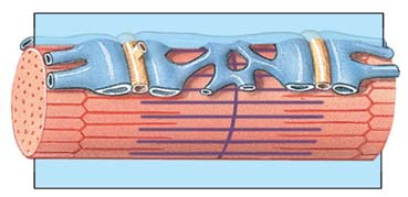
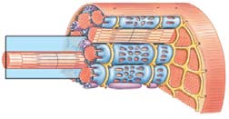
Sarcolemma: The cell membrane of a muscle fiber, surrounds the sarcoplasm (cytoplasm).
Transverse Tubules (T-tubules): Invaginations of the sarcolemma that transmit action potentials deep into the muscle fiber, ensuring simultaneous contraction.
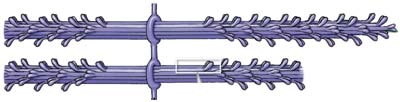
Myofibrils: Cylindrical structures composed of myofilaments (actin and myosin) responsible for contraction.
Sarcoplasmic Reticulum and Triad
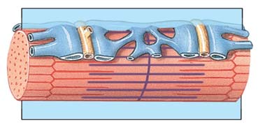
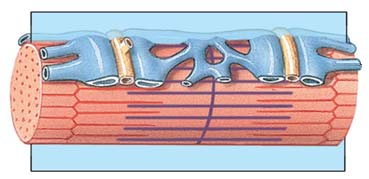
Sarcoplasmic Reticulum (SR): Specialized endoplasmic reticulum that stores and releases calcium ions (Ca2+).
Terminal Cisternae: Enlarged areas of the SR adjacent to T-tubules, concentrate and release Ca2+.
Triad: A structure formed by one T-tubule and two terminal cisternae, crucial for excitation-contraction coupling.
Sarcomere: The Contractile Unit
Sarcomere Structure
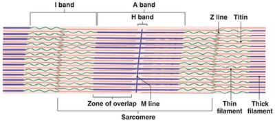
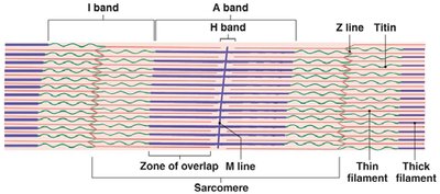
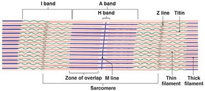
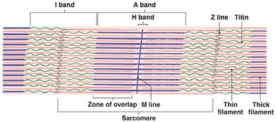
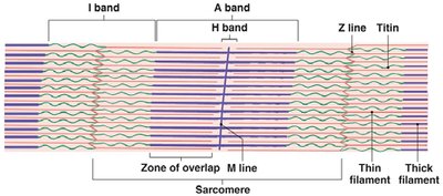
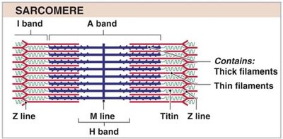
The sarcomere is the fundamental contractile unit of muscle, defined by the region between two Z lines. It contains organized thick (myosin) and thin (actin) filaments.
A Band: Dark region containing thick filaments.
I Band: Light region containing thin filaments.
H Band: Area with only thick filaments, around the M line.
M Line: Center of the A band, midline of the sarcomere.
Z Line: Boundary of the sarcomere, anchors thin filaments.
Zone of Overlap: Region where thick and thin filaments overlap, critical for contraction.
Titin: Elastic protein stabilizing thick filaments and maintaining sarcomere structure.
Muscle Striations
Striations are visible patterns in skeletal muscle caused by the arrangement of thick and thin filaments within the sarcomere.
Levels of Functional Organization
Hierarchical Structure
Skeletal muscle is organized in a hierarchical manner:
Muscle (organ): Surrounded by epimysium, contains fascicles.
Muscle Fascicle: Surrounded by perimysium, contains muscle fibers.
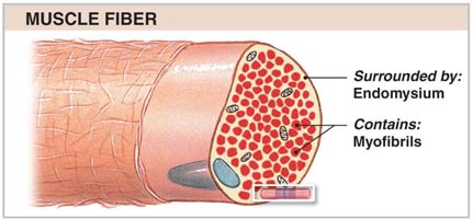
Muscle Fiber (cell): Surrounded by endomysium, contains myofibrils.
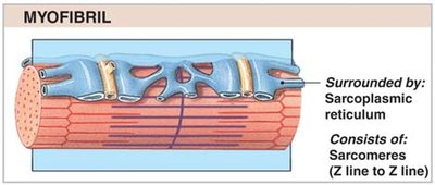
Myofibril: Surrounded by sarcoplasmic reticulum, consists of sarcomeres.
Sarcomere: Contains thick and thin filaments.
Level | Surrounding Structure | Contains |
|---|---|---|
Muscle | Epimysium | Fascicles |
Fascicle | Perimysium | Muscle fibers |
Muscle Fiber | Endomysium | Myofibrils |
Myofibril | Sarcoplasmic reticulum | Sarcomeres |
Sarcomere | None | Thick & thin filaments |
Molecular Components of Muscle Contraction
Thin Filament Proteins
F-actin: Two twisted rows of globular G-actin; active sites bind myosin.
Nebulin: Holds F-actin strands together.
Tropomyosin: Double strand, prevents actin-myosin interaction.
Troponin: Globular protein, binds tropomyosin to G-actin, controlled by Ca2+.
Initiating Contraction
Ca2+ binds to troponin, causing the troponin-tropomyosin complex to shift and expose active sites on actin.
Thick Filament Proteins
Myosin: Composed of twisted subunits; head forms cross-bridges with actin.
Titin: Provides elasticity and stabilizes thick filaments.
Sliding Filament Theory
Muscle contraction occurs as thin filaments slide past thick filaments, shortening the sarcomere and thus the muscle fiber.
The width of the A band remains constant.
Z lines move closer together.
Sliding occurs in every sarcomere along the myofibril.
Excitation-Contraction Coupling
Neuromuscular Junction
The neuromuscular junction is the site where a motor neuron stimulates a muscle fiber:
Action potential travels along the nerve axon to the synaptic terminal.
Acetylcholine (ACh) is released into the synaptic cleft.
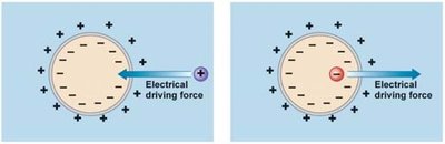
ACh binds to receptors on the sarcolemma, causing Na+ influx and generating an action potential in the muscle fiber.
ACh is broken down by acetylcholinesterase (AChE).
Excitation-Contraction Coupling
Action potential travels along T-tubules to the triad.
Ca2+ is released from the SR, triggering contraction.
Myosin heads must be "cocked" (loaded with ATP) to initiate contraction.
The Contraction Cycle
Steps of the Contraction Cycle
Exposure of active sites
Formation of cross-bridges
Pivotal movement of myosin heads
Detachment of cross-bridges
Reactivation of myosin
Fiber Shortening and Contraction Duration
As sarcomeres shorten, muscle tension is produced.
Duration depends on neural stimulus, Ca2+ availability, and ATP supply.
Relaxation Cycle
Muscle Relaxation
Nerve impulse ceases, ACh is broken down by AChE.
Ca2+ detaches from troponin and is pumped back into the SR.
Troponin-tropomyosin complex blocks actin-myosin interaction.
Cross-bridges break, muscle returns to resting length.
Muscle Cramps and Rigor Mortis
Muscle cramps: Caused by lack of ATP needed to pump Ca2+ back into SR, resulting in partial contraction.
Rigor mortis: After death, ion pumps stop, Ca2+ accumulates, and muscles remain contracted.
Summary Table: Sarcomere Bands and Lines
Band/Line | Location | Contains |
|---|---|---|
A Band | Center | Thick filaments |
I Band | Ends | Thin filaments |
H Band | Middle of A band | Thick filaments only |
M Line | Center of sarcomere | Myosin connections |
Z Line | Ends of sarcomere | Actin connections |
Zone of Overlap | Between A and I bands | Thick and thin filaments |
Key Equations
ATP Hydrolysis in Muscle Contraction
ATP is required for myosin head activation and cross-bridge cycling:
Calcium Ion Concentration
Calcium ion concentration regulates contraction:
Conclusion
Skeletal muscle tissue is highly organized, with specialized structures and molecular mechanisms that enable voluntary movement, posture, and vital physiological functions. Understanding its anatomy and physiology is fundamental for students in health and biological sciences.
