 Indietro
Ch. 10 Notes
Guida di studio - Note intelligenti
 Appunti personalizzati basati sui tuoi materiali, ampliati con definizioni chiave, esempi e contesto.
Muscle Tissue
Types and Functions of Muscle Tissue
Muscle tissue is a primary tissue responsible for contraction and movement in the human body. There are three main types of muscle tissue, each with distinct functions and properties:
Skeletal Muscle: Moves the body by pulling on bones; responsible for voluntary movements.
Cardiac Muscle: Pumps blood through the cardiovascular system; found only in the heart.
Smooth Muscle: Pushes fluids and solids through internal passageways and organs; found in walls of hollow organs.
Common properties of muscle tissue include:
Excitability (Responsiveness): Ability to receive and respond to stimuli.
Contractility: Ability of cells to shorten.
Extensibility: Ability of the muscle to stretch.
Elasticity: Ability of the muscle to recoil to its resting length.
Primary functions of skeletal muscle:
Producing movement
Maintaining posture and body position
Supporting soft tissues
Guarding body entrances and exits
Maintaining body temperature
Storing nutrients
Organization of Skeletal Muscle
Structural Hierarchy
Skeletal muscles are composed of several layers and structures that contribute to their function:
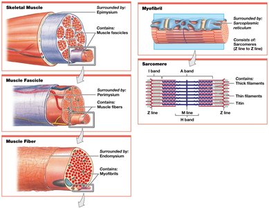
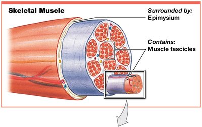
Epimysium: Surrounds the entire muscle; connected to deep fascia.
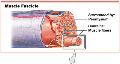
Perimysium: Surrounds individual fascicles (bundles of muscle fibers); contains blood vessels and nerves.
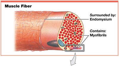
Endomysium: Surrounds individual muscle fibers; contains capillaries, nerve fibers, and myosatellite cells (stem cells).
Collagen fibers from these layers converge to form tendons (bundles) or aponeuroses (sheets), which attach muscles to bones.
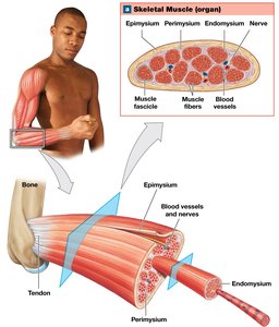
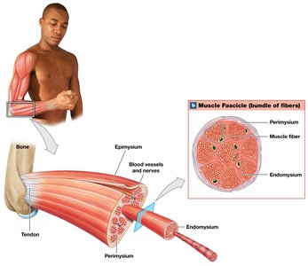
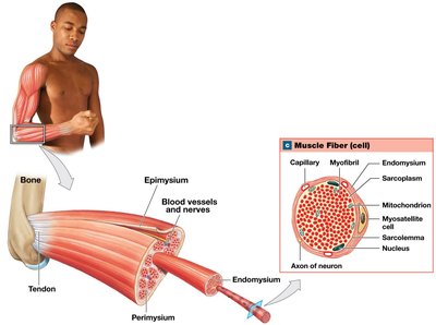
Skeletal muscles have extensive blood vessel networks for oxygen and nutrient delivery and waste removal. They contract only when stimulated by the central nervous system, making them voluntary muscles (with some exceptions like the diaphragm).
Skeletal Muscle Fibers
Development and Structure
Skeletal muscle fibers are large, multinucleate cells formed by the fusion of embryonic cells called myoblasts. These fibers are also known as striated muscle cells due to visible striations caused by the arrangement of myofibrils.
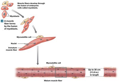
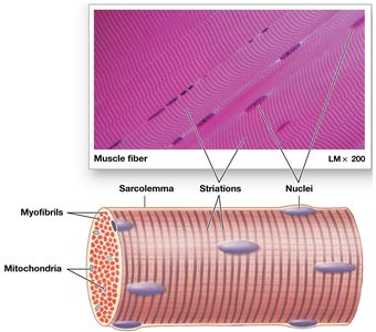
Key components of a muscle fiber:
Sarcolemma: Plasma membrane of the muscle fiber; surrounds the sarcoplasm and maintains a resting membrane potential.
Transverse Tubules (T tubules): Extensions of the sarcolemma that transmit action potentials deep into the cell.
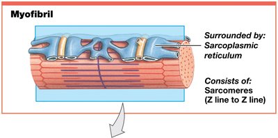
Sarcoplasmic Reticulum (SR): Tubular network specialized for calcium ion storage and release; forms terminal cisternae and triads with T tubules.
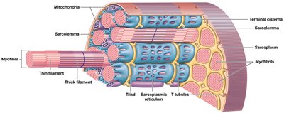
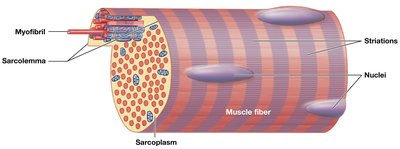
Myofibrils and Myofilaments
Myofibrils are organized collections of myofilaments responsible for muscle contraction. There are two main types of myofilaments:
Thin Filaments: Composed primarily of actin.
Thick Filaments: Composed primarily of myosin.
Sarcomere Structure
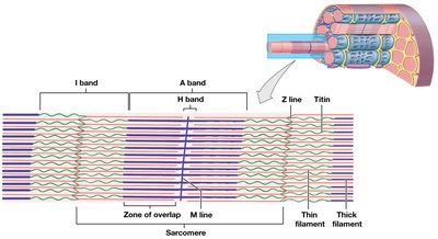
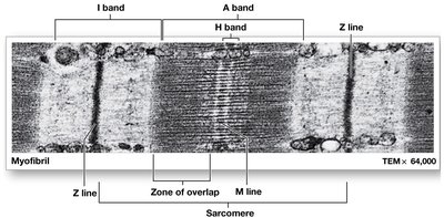
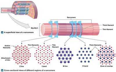
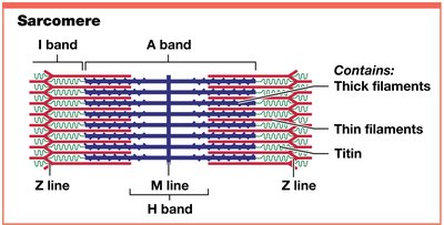
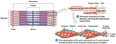
The sarcomere is the smallest contractile unit of a muscle fiber, consisting of repeating structural and functional units within myofibrils. The arrangement of thin and thick filaments within sarcomeres produces contraction.
A Bands: Dark bands containing thick filaments.
M Line: Center of A band; stabilizes thick filaments.
H Band: Region with only thick filaments.
Zone of Overlap: Area where thick and thin filaments overlap.
I Bands: Light bands containing only thin filaments.
Z Lines: Boundaries between adjacent sarcomeres.
Titin: Elastic protein that maintains alignment and restores resting length.
Thin and Thick Filament Composition
Thin filaments contain:
Tropomyosin: Covers active sites on actin, preventing actin–myosin interaction.
Troponin: Holds tropomyosin in place and binds calcium ions; moves tropomyosin off actin active sites in presence of calcium.
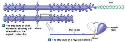
Thick filaments contain myosin:
Myosin Tail: Binds to other myosin molecules; points toward M line.
Myosin Head: Projects toward nearest thin filament; functions as ATPase.
Titin: Acts as a molecular spring, recoiling sarcomere to resting length.
Sliding-Filament Theory
During contraction, thin filaments slide toward the center of the sarcomere (M line) alongside thick filaments. This results in:
Narrowing of H and I bands
Widening of zones of overlap
Z lines moving closer together
Constant width of A band
Muscle Contraction
Excitable Membranes and Action Potentials
All cells maintain a negative resting membrane potential. In skeletal muscle cells, this is approximately -90 mV. Changes in ion distribution across the membrane can initiate contraction.
Depolarization: Membrane potential becomes less negative (influx of sodium ions).
Hyperpolarization: Membrane potential becomes more negative (outflow of potassium ions).
Repolarization: Return to resting membrane potential after depolarization.
Neurons and muscle cells can produce action potentials that propagate along the plasma membrane.
Neuromuscular Junction (NMJ)
The NMJ is the synapse between a motor neuron and a skeletal muscle fiber. Key components:
Axon Terminal: Expanded end of the motor neuron axon.
Motor End Plate: Folded membrane of the muscle fiber at the NMJ.
Synaptic Cleft: Narrow space between axon terminal and motor end plate.
Acetylcholine (ACh): Neurotransmitter released from axon terminal; binds to chemically gated Na+ channels, causing depolarization and action potential generation.
Excitation–Contraction Coupling
This process links the generation of an action potential in the sarcolemma to the start of muscle contraction:
Action potential travels down T tubules to triads.
Triggers release of calcium ions from SR.
Calcium binds to troponin, moving tropomyosin off actin active sites.
Contraction begins.
Contraction Cycle
The contraction cycle involves:
Formation of cross-bridges (myosin heads bind to actin).
Power stroke (myosin head pivots, pulling actin toward M line).
ATP is required for myosin head detachment and reactivation.
Generation of Muscle Tension
Muscle cells produce tension (pull) when they contract. Movement occurs when tension overcomes the load (resistance). All sarcomeres contract together, and the speed of shortening depends on the cycling rate (number of power strokes per second).
Relaxation
Contraction duration depends on:
Period of stimulation at NMJ
Presence of free calcium ions
Availability of ATP
When contraction ends:
ACh is broken down by acetylcholinesterase (AChE).
Calcium returns to SR via active transport.
Troponin returns to original position; tropomyosin covers actin active sites.
Rigor Mortis
After death, muscles stiffen due to ATP depletion, causing sustained contraction and inability of myosin heads to detach from actin.
Cardiac Muscle Tissue
Structure and Function
Cardiac muscle tissue is found only in the heart. Cardiac muscle cells are small, branched, striated, and have a single nucleus. They have excitable membranes, short and wide T tubules (no triads), and SR without terminal cisternae. Cardiac muscle is almost entirely dependent on aerobic metabolism and contains abundant myoglobin and mitochondria.
Intercalated discs are specialized connections joining adjacent cardiac muscle cells via gap junctions and desmosomes, allowing ions to move between cells and enabling rhythmic contractions.
Functional characteristics:
Automaticity: Ability to contract without neural stimulation (pacemaker cells).
Longer contractions and refractory periods than skeletal muscle.
Contractions require both intracellular and extracellular calcium ions.
No wave summation or tetanic contractions.
Smooth Muscle Tissue
Structure and Function
Smooth muscle tissue is found around other tissues and internal organs. It is involved in functions such as erecting hairs (integumentary system), regulating blood pressure and airflow (cardiovascular and respiratory systems), moving materials and forming sphincters (digestive and urinary systems), and transporting gametes and delivering fetuses (reproductive system).
Structural characteristics:
Long, slender, spindle-shaped cells with a single central nucleus.
No T tubules, myofibrils, or sarcomeres (non-striated).
Scattered thick filaments with many myosin heads.
Thin filaments attached to dense bodies, which connect adjacent cells and transmit contractions.
No tendons or aponeuroses.
Functional Characteristics
Excitation–Contraction Coupling: Entry of calcium ions from extracellular fluid and release from SR trigger contraction; calcium binds calmodulin, activating myosin light chain kinase.
Length–Tension Relationship: Tension and resting length are not directly related; smooth muscle can contract even when stretched (plasticity).
Control of Contractions: Multiunit smooth muscle cells are innervated in motor units; visceral smooth muscle cells are not connected to motor neurons and are controlled by pacesetter cells.
Smooth Muscle Tone: Normal background level of activity, modulated by neural, hormonal, or chemical factors.
