 Indietro
Muscle Tissue: Structure, Function, and Contraction Mechanisms
Guida di studio - Note intelligenti
 Appunti personalizzati basati sui tuoi materiali, ampliati con definizioni chiave, esempi e contesto.
Muscle Tissue Overview
Types and Functions of Muscle Tissue
Muscle tissue is a primary tissue in the human body, specialized for contraction and responsible for movement. There are three main types of muscle tissue: skeletal muscle, cardiac muscle, and smooth muscle. Each type has unique structural and functional characteristics.
Skeletal muscle: Moves the body by pulling on bones; voluntary control.
Cardiac muscle: Found only in the heart; controls heart contractions.
Smooth muscle: Controls movements inside the body, such as in blood vessels and the digestive tract.
Common properties of muscle tissue include:
Excitability (responsiveness to stimuli)
Contractility (ability to shorten)
Extensibility (ability to stretch)
Elasticity (ability to recoil)
Organization of Skeletal Muscle
Connective Tissue Layers
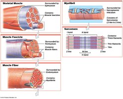
Skeletal muscles are complex organs composed of muscle tissue, connective tissues, blood vessels, and nerves. The connective tissue layers organize and protect muscle fibers:
Epimysium: Surrounds the entire muscle; separates muscle from surrounding tissues.
Perimysium: Surrounds bundles of muscle fibers called fascicles; contains blood vessels and nerves.
Endomysium: Surrounds individual muscle fibers; contains capillaries, myosatellite cells (stem cells), and nerve fibers.
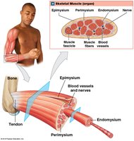
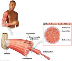
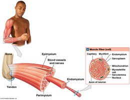
At the ends of muscles, the connective tissue layers merge to form tendons (bundles) or aponeuroses (sheets), which attach muscles to bones.
Vascular and Neural Supply
Skeletal muscles have extensive vascular networks to deliver oxygen and nutrients and remove wastes. They contract only when stimulated by the central nervous system and are considered voluntary muscles (with the exception of the diaphragm, which can function subconsciously).
Skeletal Muscle Fibers
Development and Structure
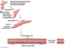
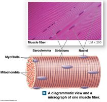
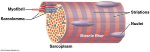
Skeletal muscle fibers are large, multinucleate cells formed by the fusion of embryonic cells called myoblasts. These fibers are also known as striated muscle cells due to their banded appearance caused by the arrangement of contractile proteins.
Membrane Systems
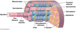
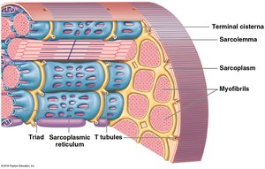
Sarcolemma: The plasma membrane of a muscle fiber; excitable and initiates contraction upon changes in membrane potential.
Sarcoplasm: The cytoplasm of a muscle fiber, containing organelles and contractile proteins.
Transverse tubules (T tubules): Invaginations of the sarcolemma that transmit action potentials deep into the cell, triggering contraction.
Sarcoplasmic reticulum (SR): Specialized endoplasmic reticulum that stores and releases calcium ions; forms terminal cisternae adjacent to T tubules, creating a triad structure.
Myofibrils and Myofilaments
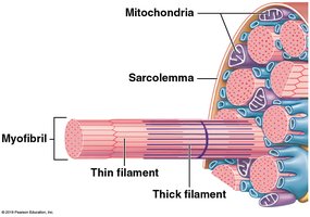
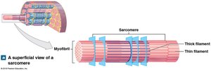
Myofibrils are cylindrical structures within muscle fibers responsible for contraction. They are composed of repeating units called sarcomeres, which contain two main types of protein filaments:
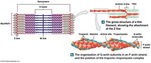
Thin filaments: Primarily actin, with associated proteins (nebulin, tropomyosin, troponin).
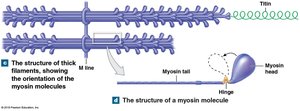
Thick filaments: Primarily myosin, with a core of titin (an elastic protein).
Sarcomere Structure and Function
Organization of the Sarcomere
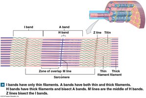
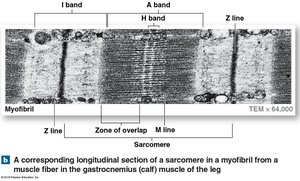
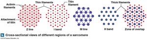
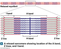
The sarcomere is the smallest functional unit of a muscle fiber. Its highly organized structure produces the striated appearance of skeletal and cardiac muscle. Key regions include:
A band: Dark region containing thick filaments (with some overlap of thin filaments).
I band: Light region containing only thin filaments.
H band: Central region of the A band with only thick filaments.
M line: Center of the A band; stabilizes thick filaments.
Z line: Boundary between adjacent sarcomeres; anchors thin filaments and titin.
Functional Organization
The arrangement of thick and thin filaments within the sarcomere allows for the sliding-filament mechanism of muscle contraction. The protein titin helps maintain alignment and restores resting length after contraction.
Thin and Thick Filaments
Thin Filaments
F-actin: Twisted strand of two rows of globular G-actin molecules; each G-actin has an active site for myosin binding.
Nebulin: Holds F-actin strands together.
Tropomyosin: Covers active sites on G-actin, preventing myosin binding in resting muscle.
Troponin: Binds to tropomyosin, G-actin, and Ca2+; regulates the position of tropomyosin.
Thick Filaments
Composed of about 300 myosin molecules, each with a tail (binds other myosin) and two heads (bind to actin).
Titin extends from the tips of thick filaments to the Z line, providing elasticity.
Sliding Filament Theory
Mechanism of Contraction
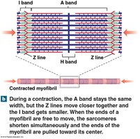
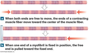
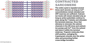
During muscle contraction, thin filaments slide toward the center of the sarcomere, causing:
Narrowing of H bands and I bands
Widening of the zone of overlap
Z lines move closer together
Width of A band remains constant
Neuromuscular Junction and Excitation-Contraction Coupling
Neuromuscular Junction (NMJ)
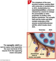
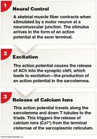
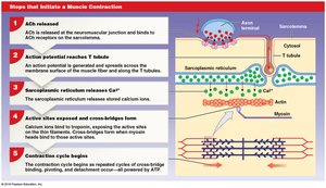
The NMJ is the synapse between a motor neuron and a skeletal muscle fiber. The process of muscle contraction is initiated by the release of the neurotransmitter acetylcholine (ACh) from the neuron, which binds to receptors on the muscle fiber, generating an action potential.
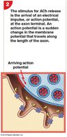
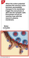
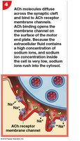
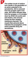
Excitation-Contraction Coupling
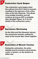
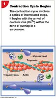
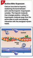
Action potentials travel along the sarcolemma and down T tubules, triggering the release of Ca2+ from the SR. Calcium binds to troponin, causing a conformational change that moves tropomyosin and exposes active sites on actin, initiating contraction.
The Contraction Cycle
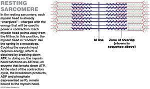
Active-site exposure
Cross-bridge formation (myosin binds actin)
Myosin head pivoting (power stroke)
Cross-bridge detachment (ATP binds myosin)
Myosin reactivation (ATP hydrolysis)
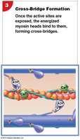
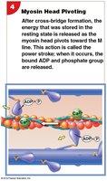
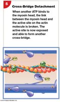
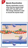
Relaxation
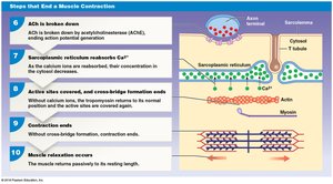
Relaxation occurs when neural stimulation ends, ACh is broken down, Ca2+ is pumped back into the SR, and active sites are re-covered by tropomyosin.
Summary Table: Skeletal Muscle Structure
Level | Surrounded by | Contains |
|---|---|---|
Skeletal Muscle | Epimysium | Muscle fascicles |
Muscle Fascicle | Perimysium | Muscle fibers |
Muscle Fiber | Endomysium | Myofibrils |
Myofibril | Sarcoplasmic reticulum | Sarcomeres |
Sarcomere | None (functional unit) | Thick and thin filaments, titin |
Additional info: This summary covers the structure, organization, and contraction mechanisms of skeletal muscle tissue, integrating key diagrams for visual reinforcement. For a complete understanding, students should also study the regulation of contraction, energy metabolism, and differences between muscle tissue types.
