 Indietro
Muscle Tissue: Structure, Function, and Contraction
Guida di studio - Note intelligenti
 Appunti personalizzati basati sui tuoi materiali, ampliati con definizioni chiave, esempi e contesto.
Muscle Tissue
Types of Muscle Tissue
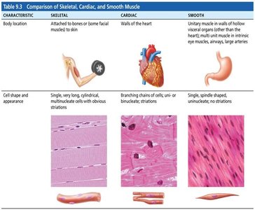
Muscle tissue is specialized for contraction and is essential for movement, posture, and various bodily functions. There are three main types of muscle tissue: skeletal, cardiac, and smooth muscle. Each type has distinct structural and functional characteristics.
Skeletal Muscle: Attached to bones and skin, responsible for voluntary movements. Cells are long, cylindrical, multinucleated, and striated.
Cardiac Muscle: Found only in the heart, responsible for pumping blood. Cells are branching, striated, usually uninucleate, and involuntary.
Smooth Muscle: Located in walls of hollow organs (e.g., stomach, blood vessels). Cells are spindle-shaped, uninucleate, non-striated, and involuntary.
Key Characteristics Shared by All Muscle Types:
Excitability: Ability to respond to stimuli.
Contractility: Ability to shorten forcibly.
Extensibility: Ability to be stretched.
Elasticity: Ability to recoil to resting length.
Connective Tissue Components of Skeletal Muscle
Organization and Structure
Skeletal muscle is an organ composed of muscle fibers, connective tissue, blood vessels, and nerves. The connective tissue sheaths support and protect muscle fibers and contribute to force transmission.
Epimysium: Surrounds the entire muscle.
Perimysium: Surrounds bundles of muscle fibers called fascicles.
Endomysium: Surrounds individual muscle fibers.
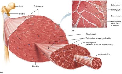
Muscles attach to bones via tendons (rope-like) or aponeuroses (sheet-like). Attachments can be direct (muscle to bone) or indirect (via connective tissue).
Structural Parts of Skeletal Muscle Tissue
Muscle Fiber Anatomy
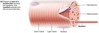
Skeletal muscle fibers are long, cylindrical cells with multiple nuclei. They contain specialized structures for contraction:
Sarcolemma: Plasma membrane of the muscle fiber.
Sarcoplasm: Cytoplasm containing glycosomes (glycogen storage) and myoglobin (oxygen storage).
Myofibrils: Rod-like structures that run the length of the cell and contain the contractile elements.
Myofibrils and Sarcomeres
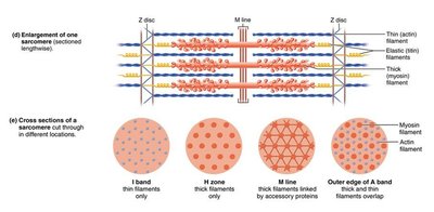
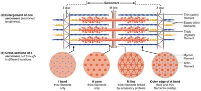
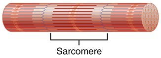
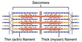
Myofibrils are composed of repeating units called sarcomeres, the functional units of muscle contraction. Sarcomeres are defined by Z discs and contain thick (myosin) and thin (actin) filaments arranged in a precise pattern, producing the striated appearance of skeletal muscle.
Striations: Alternating dark (A bands) and light (I bands) regions due to the arrangement of myofilaments.
Sarcomere: Extends from one Z disc to the next; contains A band (thick filaments) and I band (thin filaments).
Myofilaments: Thick and Thin Filaments
Myofilaments are the contractile proteins within the sarcomere:
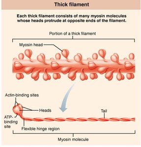
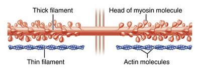
Thick Filaments: Composed of myosin molecules, each with a head (binds actin and ATP) and a tail.
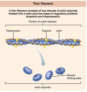
Thin Filaments: Composed of actin, tropomyosin, and troponin. Actin has binding sites for myosin heads; tropomyosin and troponin regulate interaction.
Sarcomere Organization
The arrangement of thick and thin filaments within the sarcomere is critical for muscle contraction. The sliding of thin filaments past thick filaments shortens the sarcomere, resulting in muscle contraction.
Major Events of Muscle Contraction and Relaxation
Neuromuscular Junction and Excitation
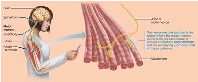
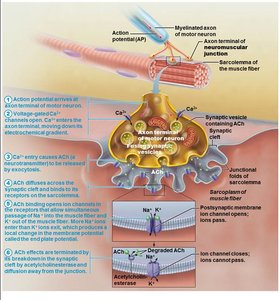
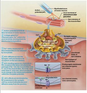
Muscle contraction is initiated by signals from motor neurons. The neuromuscular junction (NMJ) is the site where the motor neuron communicates with the muscle fiber.
Motor Neuron: Sends action potential to muscle fiber.
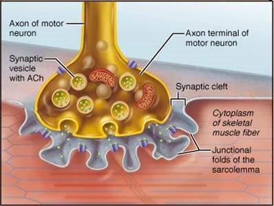
Axon Terminal: Releases acetylcholine (ACh) into the synaptic cleft.
Junctional Folds: Contain ACh receptors on the sarcolemma.
Events at the Neuromuscular Junction
Action potential arrives at axon terminal.
Voltage-gated Ca2+ channels open; Ca2+ enters terminal.
Ca2+ triggers release of ACh into synaptic cleft.
ACh binds to receptors, opening Na+ channels and generating an end plate potential.
Acetylcholinesterase degrades ACh, terminating the signal.
Action Potential and Muscle Fiber Response
Depolarization: Na+ influx causes local depolarization (end plate potential). If threshold is reached, an action potential is generated and propagated along the sarcolemma.
Repolarization: K+ efflux restores resting membrane potential. The Na+-K+ pump reestablishes ionic conditions.
Energy Sources for Muscle Contraction
ATP and Muscle Contraction
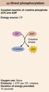
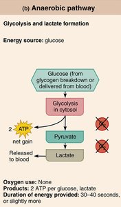
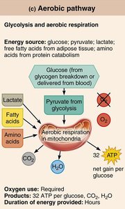
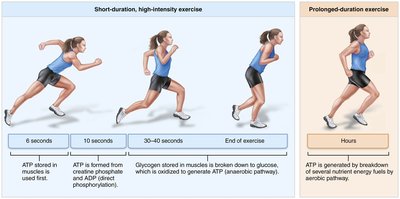
ATP is the immediate source of energy for muscle contraction. It is required for cross-bridge cycling, Ca2+ pumping, and restoring ion gradients. Because ATP stores are limited, muscle fibers regenerate ATP through three main pathways:
Direct Phosphorylation: Creatine phosphate (CP) donates a phosphate to ADP to form ATP. Provides energy for short bursts (about 15 seconds).
Anaerobic Pathway: Glycolysis breaks down glucose to pyruvate, producing ATP and lactic acid (when oxygen is limited). Supports moderate activity for 30–40 seconds.
Aerobic Pathway: Aerobic respiration in mitochondria uses glucose, fatty acids, or amino acids to produce ATP, CO2, and H2O. Supports prolonged activity.
The "All or None" Principle
Action Potential and Muscle Response
The "all or none" principle states that once a muscle fiber or neuron is stimulated to threshold, it will generate a full action potential and contract maximally. There are no partial action potentials; the response is always complete if threshold is reached.
Analogy: Like pulling a trigger on a gun—either it fires or it does not, regardless of how hard the trigger is pulled beyond the threshold.
Types of Skeletal Muscle Contraction
Isometric and Isotonic Contractions
Muscle contractions can be classified based on whether the muscle changes length during contraction:
Isometric Contraction: Muscle develops tension but does not change length (e.g., holding a weight steady).
Isotonic Contraction: Muscle changes length to move a load. Subdivided into:
Concentric: Muscle shortens as it contracts (e.g., lifting a book).
Eccentric: Muscle lengthens while maintaining tension (e.g., lowering a book).
Clinical Implications
Muscle Disorders
Duchenne Muscular Dystrophy (DMD): Genetic disorder causing defective dystrophin, leading to muscle degeneration.
Myasthenia Gravis: Autoimmune disease where ACh receptors are destroyed, causing muscle weakness.
Rigor Mortis: Postmortem muscle stiffness due to lack of ATP and sustained cross-bridge formation.
Disuse Atrophy: Muscle wasting due to inactivity or loss of neural stimulation.
