 Indietro
Muscle Tissue: Structure, Function, and Contraction Mechanisms
Guida di studio - Note intelligenti
 Appunti personalizzati basati sui tuoi materiali, ampliati con definizioni chiave, esempi e contesto.
Muscle Tissue and Homeostasis
Overview of Muscle Tissue Functions
Muscle tissue is essential for maintaining homeostasis in the human body. It produces body movements, moves substances within the body, and aids in the maintenance of body temperature by generating heat as a byproduct. The scientific study of muscles is known as myology.
Body Movements: Muscles contract to produce voluntary and involuntary movements.
Movement of Substances: Muscles move blood, food, and other substances through various systems.
Heat Production: Muscle contraction generates heat, helping regulate body temperature.
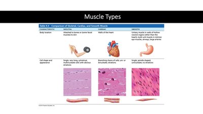
Muscle Types
Classification and Comparison of Muscle Tissue
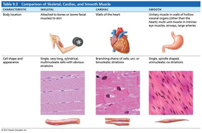
There are three main types of muscle tissue: skeletal, cardiac, and smooth. Each type has distinct structural and functional characteristics.
Skeletal Muscle: Attached to bones (and some to skin), striated, voluntary, multinucleated.
Cardiac Muscle: Found only in the heart, striated, involuntary, branched cells.
Smooth Muscle: Located in walls of hollow organs and blood vessels, non-striated, involuntary, spindle-shaped cells.
Characteristic | Skeletal | Cardiac | Smooth |
|---|---|---|---|
Body Location | Attached to bones or skin | Walls of the heart | Walls of hollow organs, blood vessels |
Cell Shape & Appearance | Long, cylindrical, multinucleated, striated | Branched, uni- or binucleated, striated | Spindle-shaped, uninucleated, no striations |
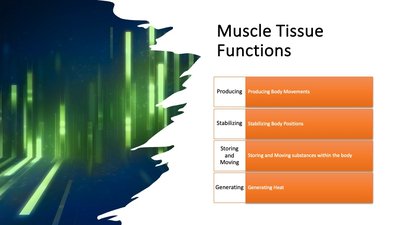
Muscle Tissue Functions
Key Functions of Muscle Tissue
Muscle tissue performs several vital functions in the body:
Producing Body Movements: Enables locomotion and manipulation of the environment.
Stabilizing Body Positions: Maintains posture and joint stability.
Storing and Moving Substances: Sphincters control passage of substances; smooth muscle moves food and blood. skeletal also helps with this
Generating Heat: Thermogenesis during contraction helps maintain body temperature.
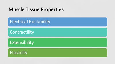
Muscle Tissue Properties
Unique Properties of Muscle Tissue
action potential=voltage. can be autorythmic or chemical (pH)
Muscle tissue exhibits four key properties that enable its function:
Electrical Excitability: Ability to respond to stimuli by producing action potentials.
Contractility: Ability to contract(get short and extend) forcefully when stimulated.
Extensibility: Ability to stretch without being damaged. connective tissue limits the extension of muscles
Elasticity: Ability to return to original shape after contraction or extension. need to have ability to return to original shape
Skeletal Muscle Tissue – Connective Tissue Components
Organization and Structure of Skeletal Muscle
Skeletal muscle is an organ composed of muscle fibers, blood vessels, nerves, and connective tissue. The connective tissue layers include:
Epimysium: Surrounds the entire muscle.
Perimysium: Surrounds bundles of muscle fibers (fascicles).
Endomysium: Surrounds individual muscle fibers.
Tendon: Connects muscle to bone.
Aponeurosis: Flat sheet for muscle attachment.
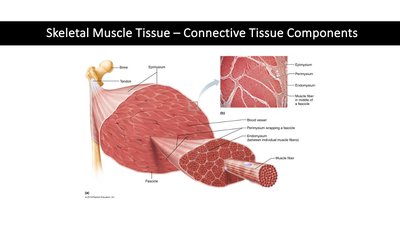
muscle organs
Microscopic Anatomy of Skeletal Muscle Fibers
Structure of Muscle Fibers
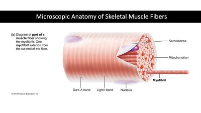
Muscle fibers are long, cylindrical cells containing multiple nuclei and specialized organelles:
Sarcolemma: Plasma membrane of muscle fiber. plasmalemma
Sarcoplasm: Cytoplasm of muscle fiber.
Sarcoplasmic Reticulum (SR): Stores and releases calcium ions. endoplasmic reticulum
Myofibrils: Contractile organelles composed of sarcomeres.
Myoglobin: Oxygen-binding protein for ATP synthesis.
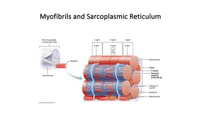
Myofibrils and Sarcoplasmic Reticulum
Organization of Contractile Structures
Myofibrils are composed of repeating units called sarcomeres, which are the basic functional units of muscle contraction. The sarcoplasmic reticulum surrounds myofibrils and releases calcium ions to initiate contraction.
Transverse Tubules (T-Tubules): Invaginations of the sarcolemma that conduct action potentials.
Triad: Structure formed by a T-tubule and two terminal cisterns of the SR.
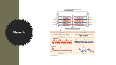
Filaments
Types of Myofilaments
Myofibrils contain two main types of filaments:
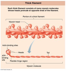
Thick Filaments: Composed mainly of myosin.
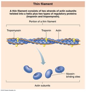
Thin Filaments: Composed mainly of actin, with regulatory proteins troponin and tropomyosin.
Filament Type | Main Protein | Associated Proteins |
|---|---|---|
Thick | Myosin | None |
Thin | Actin | Troponin, Tropomyosin |
Sarcomere
Structure and Functional Zones
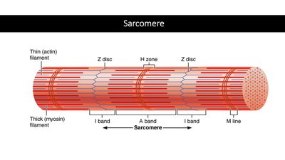
The sarcomere is the basic contractile unit of muscle fiber, defined by Z-discs. It contains distinct regions:
A Band: Dark region with thick filaments.
I Band: Light region with thin filaments.
H Zone: Area with only thick filaments.
M Line: Center of the H zone, supporting thick filaments.
Z Discs: Boundaries of the sarcomere.
Muscle Proteins
Contractile, Regulatory, and Structural Proteins
Muscle contraction depends on three classes of proteins:
Contractile Proteins: Actin (thin filament) and myosin (thick filament) generate force. for contraction
Regulatory Proteins: Tropomyosin and troponin control contraction by covering or exposing myosin-binding sites. on/off switch
Structural Proteins: Titin, α-actinin, myomesin, nebulin, and dystrophin maintain alignment and elasticity.
titin=largest protein in the body (35,000 ish amino acids in polypeptide chain) responsible for elasticity in muscle cells
alpha-actinin= think line protein that attaches actin molecules to thin filament
nebulin= stretches across entire thin filament in sarcomere, attaches thin filaments to z discs
myomesin= m line protein, attaches thick filaments to thick filaments
dystrophin= stabalizes or reinforces sarcolemma, transfers muscle contraction tension to tendons
Quick Check: Muscle Structure Matching
Matching Descriptions to Muscle Components
Item | Description |
|---|---|
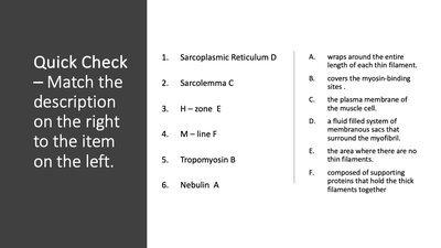
Sarcoplasmic Reticulum | A fluid filled system of membranous sacs that surround the myofibril. calcium storage |
Sarcolemma | The plasma membrane of the muscle cell. |
H – zone | The area where there are no thin filaments. |
M – line | Composed of supporting proteins that hold the thick filaments together. |
Tropomyosin | Covers the myosin-binding sites. |
Nebulin | Wraps around the entire length of each thin filament. |
How Muscle Contractions Happen: Overview
Sequence of Muscle Contraction
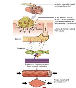
Muscle contraction is initiated by an action potential, leading to the release of calcium ions, which bind to troponin and allow myosin to interact with actin. This process shortens the muscle fiber and produces tension.
Action Potential: Arrives at neuromuscular junction.
Calcium Release: Calcium binds to troponin, moving tropomyosin.
Cross-Bridge Formation: Myosin binds to actin.
Contraction: Muscle fiber shortens.
The Contraction Cycle
Steps of the Cross-Bridge Cycle
The contraction cycle consists of four steps:
ATP Hydrolysis: Myosin head hydrolyzes ATP, becoming energized.
Attachment: Myosin head binds to actin, forming a cross-bridge.
Power Stroke: Myosin head pivots, pulling actin toward the center.
Detachment: Myosin releases from actin upon binding new ATP.
Additional info:
These notes cover the structure and function of muscle tissue, including microscopic anatomy, contractile mechanisms, and the molecular basis of muscle contraction. The content is directly relevant to Anatomy & Physiology college courses, specifically chapters on muscle tissue and the muscular system.
rigor mortis- q on final- stiffness after death, proves we need atp for certain things to happen in muscles. dead people don't produce atp, so active transport cannot transport calcium by atp, if a new atp doesnt bind to the myosin head it stays attached to actin, meaning muscles become very stiff/rigid
