 Indietro
Muscle Tissue: Structure, Function, and Contraction
Guida di studio - Note intelligenti
 Appunti personalizzati basati sui tuoi materiali, ampliati con definizioni chiave, esempi e contesto.
Muscle Tissue
Types of Muscle Tissue
Muscle tissue is essential for movement and force generation in the human body. There are three primary types of muscle tissue: skeletal, cardiac, and smooth. Each type has distinct structural and functional characteristics.
Characteristic | Skeletal | Cardiac | Smooth |
|---|---|---|---|
Body location | Attached to bones or skin | Walls of the heart | Walls of hollow organs (e.g., stomach, bladder, arteries) |
Cell shape and appearance | Single, very long, cylindrical, multinucleate cells with obvious striations | Branching chains of cells; uni- or binucleate; striations | Single, spindle-shaped, uninucleate; no striations |
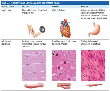
Skeletal muscle: Voluntary, striated, rapid contractions, tires easily, powerful.
Cardiac muscle: Involuntary, striated, contracts at a steady rate, regulated by pacemaker and nervous system.
Smooth muscle: Involuntary, non-striated, found in walls of hollow organs, slow and sustained contractions.
Characteristics and Functions of Muscle Tissue
All muscle tissues share four main characteristics:
Excitability: Ability to respond to stimuli.
Contractility: Ability to shorten forcibly when stimulated.
Extensibility: Ability to be stretched.
Elasticity: Ability to recoil to resting length.
Four important functions of muscle tissue:
Produce movement (locomotion, manipulation, digestion, blood pumping)
Maintain posture and body position
Stabilize joints
Generate heat as they contract
Structure of Skeletal Muscle Tissue
Connective Tissue Sheaths
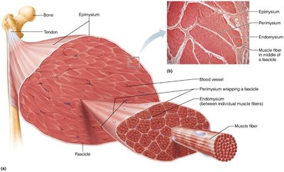
Skeletal muscle is composed of multiple tissues, including nerve and blood supply, connective tissue sheaths, and attachments. Each muscle and muscle fiber is covered by connective tissue sheaths that support and reinforce the muscle.
Epimysium: Surrounds the entire muscle.
Perimysium: Surrounds groups of muscle fibers (fascicles).
Endomysium: Surrounds individual muscle fibers.
Muscle Fiber Structure
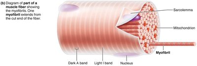
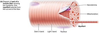
Skeletal muscle fibers are long, cylindrical cells with multiple nuclei. They contain specialized organelles for contraction and energy storage.
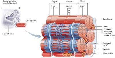
Sarcolemma: Plasma membrane of the muscle fiber.
Sarcoplasm: Cytoplasm of the muscle fiber, rich in glycogen and myoglobin.
Myofibrils: Densely packed, rodlike elements responsible for contraction.
Striations and Sarcomeres
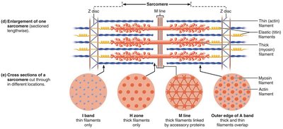
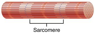
Striations are formed by repeating series of dark and light bands along the length of each myofibril. The sarcomere is the smallest contractile unit of muscle fiber, aligned end to end along myofibrils.
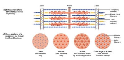
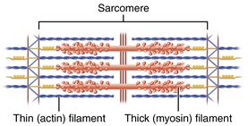
Sarcomere: Functional unit of muscle fiber, composed of myofilaments.
Myofilaments: Thick (myosin) and thin (actin) filaments arranged within the sarcomere.
Myofilaments: Actin and Myosin
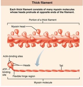
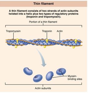
Myofilaments are organized within the sarcomere to facilitate contraction. Actin and myosin are the primary contractile proteins.
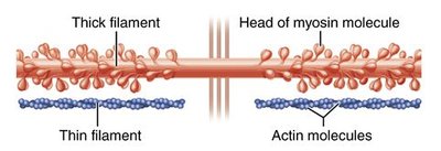
Actin (thin filament): Contains binding sites for myosin heads; associated with regulatory proteins tropomyosin and troponin.
Myosin (thick filament): Has heads that bind to actin during contraction, forming cross bridges.
Levels of Muscle Organization
Muscle structure is organized hierarchically:
Myofibril: Complex organelle composed of bundles of myofilaments.
Sarcomere: Segment of a myofibril, contractile unit.
Myofilament: Extended macromolecular structure, thick (myosin) and thin (actin) filaments.
Clinical Implications: Duchenne Muscular Dystrophy (DMD)
DMD is caused by a defective gene for dystrophin, a protein that links thin filaments to the extracellular matrix and stabilizes the sarcolemma. The sarcolemma tears easily, allowing excess calcium entry, damaging contractile fibers, and leading to muscle cell apoptosis and loss of muscle mass.

Sarcoplasmic Reticulum and T Tubules
The sarcoplasmic reticulum (SR) is a network of smooth endoplasmic reticulum tubules surrounding each myofibril, regulating intracellular Ca2+ levels. T tubules are extensions of the sarcolemma that increase surface area and allow electrical impulses to reach deep into the muscle fiber, triggering calcium release from the SR.
Muscle Contraction and Relaxation
Sliding Filament Model
During contraction, thin filaments slide past thick filaments, causing actin and myosin to overlap more. Neither filament changes length; instead, the overlap increases.
Neuromuscular Junction and Action Potential
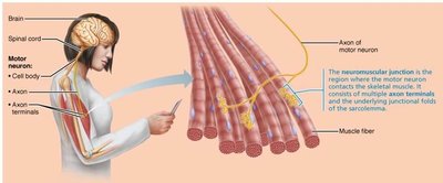
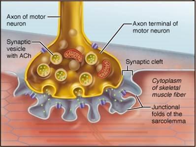
Skeletal muscles are stimulated by somatic motor neurons. The neuromuscular junction is the site where the motor neuron contacts the muscle fiber, consisting of axon terminals, synaptic cleft, and junctional folds.
Events at the Neuromuscular Junction
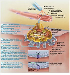
Action potential arrives at axon terminal.
Voltage-gated calcium channels open; calcium enters motor neuron.
Calcium entry causes release of ACh into synaptic cleft.
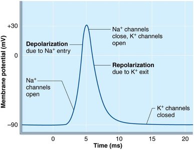
ACh binds to receptors on sarcolemma, opening Na+ channels.
Na+ entry causes end plate potential (local depolarization).
Acetylcholinesterase degrades ACh.
Repolarization
Repolarization restores resting conditions after an action potential. The Na+-K+ pump returns ions to their original locations, and the muscle fiber enters a refractory period during which it cannot be stimulated.
Clinical Implications: Myasthenia Gravis and Rigor Mortis
Myasthenia gravis: Autoimmune disease where ACh receptors are destroyed, causing muscle weakness.
Rigor mortis: After death, ATP is depleted, calcium accumulates, and muscles remain contracted until proteins break down.
Energy Sources for Muscle Contraction
ATP and Its Regeneration
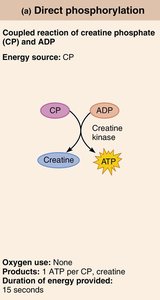
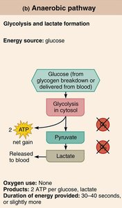
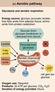
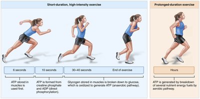
ATP is the only energy source for contractile activities and must be regenerated quickly. Muscle fibers use three mechanisms to regenerate ATP:
Direct phosphorylation of ADP by creatine phosphate (CP)
Anaerobic pathway: Glycolysis and lactate formation
Aerobic pathway: Glycolysis and aerobic respiration in mitochondria
Principles of Muscle Contraction
All-or-None Principle
The action potential is always a full response; it is an all-or-nothing process. If the stimulus is strong enough, the neuron fires, and the muscle contracts fully. This ensures reliable transmission of information.
Types of Skeletal Muscle Contraction
Isometric contraction: Muscle tension increases but does not exceed load; muscle does not shorten.
Isotonic contraction: Muscle shortens because tension exceeds load. Can be concentric (muscle shortens and does work) or eccentric (muscle lengthens while generating force).
Clinical Implications: Muscle Health
Disuse Atrophy
Muscles must be active to remain healthy. Disuse atrophy occurs due to immobilization or loss of neural stimulation, leading to degeneration and loss of muscle mass. Rehabilitation becomes impossible if muscle tissue is replaced by fibrous connective tissue.
Summary Table: Muscle Tissue Comparison
Type | Location | Striations | Control | Cell Shape |
|---|---|---|---|---|
Skeletal | Bones/skin | Striated | Voluntary | Long, cylindrical, multinucleate |
Cardiac | Heart | Striated | Involuntary | Branched, uni/binucleate |
Smooth | Hollow organs | Non-striated | Involuntary | Spindle-shaped, uninucleate |
