 Indietro
Muscle Tissue: Structure, Function, and Contraction
Guida di studio - Note intelligenti
 Appunti personalizzati basati sui tuoi materiali, ampliati con definizioni chiave, esempi e contesto.
Muscle Tissue
Types of Muscle Tissue
Muscle tissue is essential for movement and force generation in the human body. There are three primary types of muscle tissue: skeletal, cardiac, and smooth. Each type has distinct structural and functional characteristics.
Skeletal muscle: Attached to bones and skin, responsible for voluntary movements. Cells are long, cylindrical, multinucleated, and striated.
Cardiac muscle: Found in the walls of the heart, responsible for involuntary contractions. Cells are branching, striated, and usually uninucleate or binucleate.
Smooth muscle: Located in walls of hollow organs (e.g., stomach, bladder, arteries), responsible for involuntary movements. Cells are spindle-shaped, uninucleate, and non-striated.
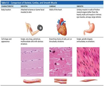
Characteristics and Functions of Muscle Tissue
All muscle tissues share four main characteristics:
Excitability: Ability to respond to stimuli.
Contractility: Ability to shorten forcibly when stimulated.
Extensibility: Ability to be stretched.
Elasticity: Ability to recoil to resting length.
Muscle tissue functions include producing movement, maintaining posture, stabilizing joints, and generating heat.
Structure of Skeletal Muscle Tissue
Connective Tissue Sheaths
Skeletal muscle is organized into layers of connective tissue that support and reinforce the muscle:
Epimysium: Surrounds the entire muscle.
Perimysium: Surrounds groups of muscle fibers (fascicles).
Endomysium: Surrounds individual muscle fibers.
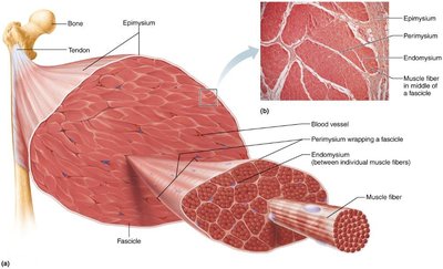
Muscle Fiber Structure
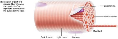
Skeletal muscle fibers are long, cylindrical cells with multiple nuclei. Key components include:
Sarcolemma: Plasma membrane of the muscle fiber.
Sarcoplasm: Cytoplasm of the muscle fiber, containing glycosomes (glycogen storage) and myoglobin (oxygen storage).
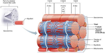
Myofibrils: Densely packed, rodlike elements responsible for contraction.
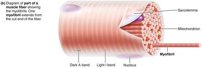
Striations and Sarcomeres
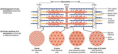
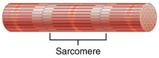
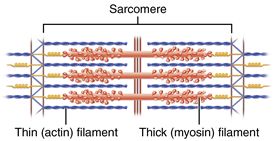
Striations are visible bands formed by the arrangement of myofibrils. The sarcomere is the smallest contractile unit of muscle fiber, composed of thick (myosin) and thin (actin) filaments.
Sarcomeres: Align end-to-end along myofibrils, like boxcars in a train.
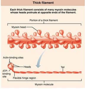
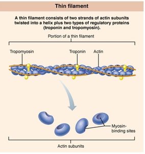
Myofilaments: Actin (thin) and myosin (thick) filaments are organized within the sarcomere.
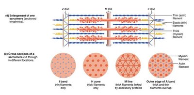
Myofilament Structure
Myofilaments are the contractile proteins within the sarcomere:
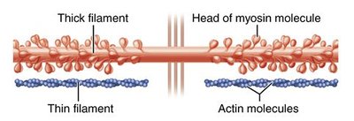
Thick filaments: Composed of myosin molecules, each with a head that binds to actin and an ATP-binding site.
Thin filaments: Composed of actin, tropomyosin, and troponin. Actin provides binding sites for myosin heads.
Sarcoplasmic Reticulum and T Tubules
The sarcoplasmic reticulum (SR) is a network of smooth endoplasmic reticulum that regulates intracellular calcium levels. T tubules are extensions of the sarcolemma that allow electrical impulses to reach deep into the muscle fiber, triggering calcium release from the SR.
Muscle Contraction and Relaxation
Sliding Filament Model
Muscle contraction occurs when thin filaments slide past thick filaments, increasing overlap. Myosin heads bind to actin, forming cross bridges, and pull the filaments together.
Neuromuscular Junction and Action Potential
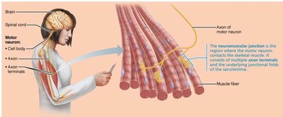
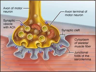
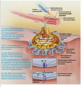
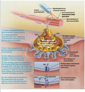
The neuromuscular junction is where the motor neuron communicates with the muscle fiber. The process involves:
Action potential arrives at axon terminal.
Voltage-gated calcium channels open; calcium enters the neuron.
Calcium triggers release of acetylcholine (ACh) into the synaptic cleft.
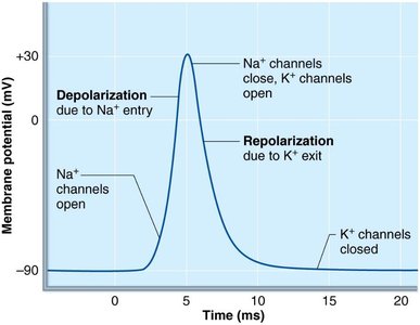
ACh binds to receptors on the sarcolemma, opening Na+ channels.
Na+ influx causes depolarization (end plate potential).
Acetylcholinesterase degrades ACh, ending the signal.
Depolarization and Repolarization
Depolarization occurs when Na+ enters the cell, making the interior more positive. If threshold is reached, an action potential is generated and spreads across the sarcolemma. Repolarization restores resting conditions via the Na+-K+ pump.
Energy Sources for Muscle Contraction
ATP and Regeneration Mechanisms
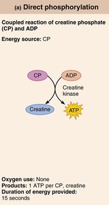
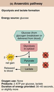
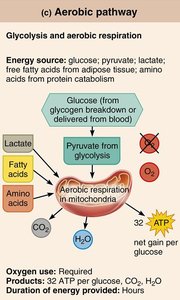
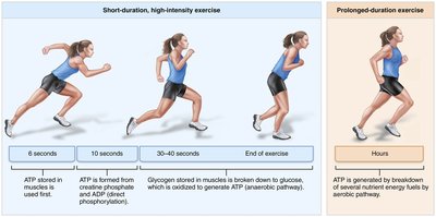
ATP is the primary energy source for muscle contraction. It is regenerated by:
Direct phosphorylation: Creatine phosphate (CP) donates a phosphate to ADP, forming ATP.
Anaerobic pathway: Glycolysis produces ATP and lactate without oxygen.
Aerobic pathway: Glycolysis followed by aerobic respiration in mitochondria produces more ATP with oxygen.
Principles and Types of Muscle Contraction
All-or-None Principle
The action potential in muscle fibers is an all-or-nothing response. If the stimulus reaches threshold, a full action potential is generated; otherwise, no response occurs. This ensures reliable signal transmission.
Types of Skeletal Muscle Contraction
Muscle contraction produces tension, which may or may not result in muscle shortening:
Isometric contraction: Muscle tension increases but does not exceed the load; muscle does not shorten.
Isotonic contraction: Muscle tension exceeds the load and muscle shortens.
Concentric: Muscle shortens and does work (e.g., lifting a book).
Eccentric: Muscle lengthens while generating force (e.g., lowering a book).
Clinical Implications
Duchenne Muscular Dystrophy (DMD)
DMD is caused by a defective gene for dystrophin, leading to muscle fiber damage, inflammation, and loss of muscle mass.
Myasthenia Gravis
An autoimmune disease where ACh receptors are destroyed, resulting in muscle weakness.
Rigor Mortis
After death, ATP production ceases, calcium accumulates, and muscles remain contracted until proteins break down.
Disuse Atrophy
Muscles lose mass and strength when not used, and rehabilitation becomes impossible if muscle tissue is replaced by fibrous connective tissue.
Structure | Description |
|---|---|
Myofibril | Rodlike contractile elements composed of sarcomeres; appear banded. |
Sarcomere | Contractile unit made of myofilaments (actin and myosin). |
Myofilament | Thick (myosin) and thin (actin) filaments; sliding produces muscle shortening. |
Type | Location | Striated/Non-striated | Voluntary/Involuntary | Cell Shape |
|---|---|---|---|---|
Skeletal | Bones/skin | Striated | Voluntary | Long, cylindrical, multinucleate |
Cardiac | Heart | Striated | Involuntary | Branching, uninucleate/binucleate |
Smooth | Hollow organs | Non-striated | Involuntary | Spindle-shaped, uninucleate |
Example: Biceps muscle contracts concentrically to lift a book and eccentrically to lower it.
Additional info: Tables above are inferred from the original content and images for clarity and completeness.
