 Indietro
Muscle Tissue: Structure, Function, and Physiology
Guida di studio - Note intelligenti
 Appunti personalizzati basati sui tuoi materiali, ampliati con definizioni chiave, esempi e contesto.
Muscle Tissue
Introduction
Muscle tissue is essential for movement, posture, stabilization, and heat production in the human body. The study of muscles is known as myology. Muscles contract and relax to produce motion, with the skeletal system providing leverage and support.
Properties of Muscle Tissue
Excitability: Ability to respond to chemical signals from nerve cells.
Conductivity: Ability to propagate electrical signals across the cell membrane.
Contractility: Ability to shorten and generate force.
Extensibility: Ability to stretch without damage.
Elasticity: Ability to return to original shape after stretching.
Functions of Muscle Tissue
Motion: Includes both external (walking, running, talking) and internal (heartbeat, digestion) movements.
Posture: Maintains body posture.
Stabilization: Stabilizes joints, with muscle tone even at rest.
Thermogenesis: Generates heat through contractions.
Connective Tissue Wrappings of Skeletal Muscle Tissue
Organization and Structure
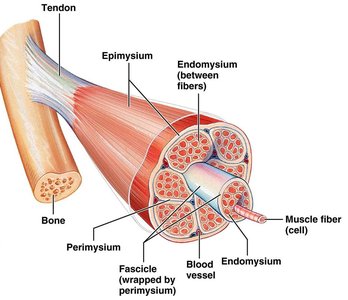
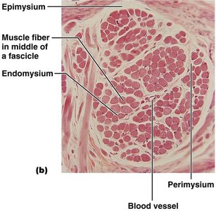
Skeletal muscle is organized into layers of connective tissue that provide support and compartmentalization:
Epimysium: Wraps the entire muscle.
Perimysium: Subdivides muscle into fascicles (bundles of 10-100 muscle fibers).
Endomysium: Wraps individual muscle fibers.
Nerve and Blood Supply
Each muscle fiber is innervated by a branch of a motor nerve.
Each muscle receives its own arteries and veins.
Blood vessels branch extensively to supply each muscle fiber directly.
Types of Muscle Tissue
Overview
There are three main types of muscle tissue, each with distinct characteristics and functions:
Skeletal muscle: Striated, voluntary, attached to bones.
Cardiac muscle: Striated, involuntary, forms the heart wall.
Smooth (visceral) muscle: Nonstriated, involuntary, found in internal organs.
Skeletal Muscle
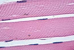
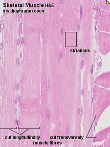
Skeletal muscle is responsible for voluntary movements and is characterized by its striated appearance and multinucleated, long, cylindrical cells.
Attached primarily to bones.
Voluntary control.
Contracts quickly, fatigues easily.
Can generate varying forces.
Striated and multinucleated.
Cardiac Muscle
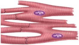
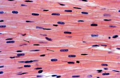
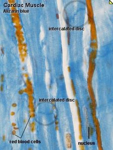
Cardiac muscle forms the myocardium of the heart and contracts involuntarily. It is striated, branched, and contains intercalated discs for synchronized contraction.
Involuntary control.
Contracts without external stimuli (autorhythmic).
Fast contraction, non-fatigable.
Beats at a constant rhythm, modifiable by neural and hormonal signals.
Branched, striated, uninucleated cells.
Intercalated discs present.
Smooth Muscle
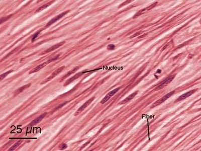
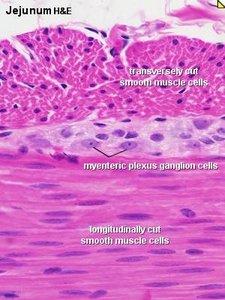
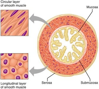
Smooth muscle is found in the walls of hollow organs and blood vessels. It is nonstriated, involuntary, and contracts slowly but is resistant to fatigue.
Located in digestive, respiratory, reproductive tracts, and blood vessels.
Involuntary control.
Long, slow contractions, non-fatigable.
Nonstriated, uninucleated.
Microscopic Anatomy of Skeletal Muscle Fiber
Muscle Fiber Structure
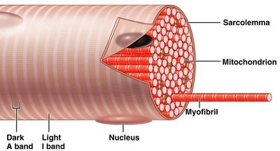
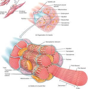
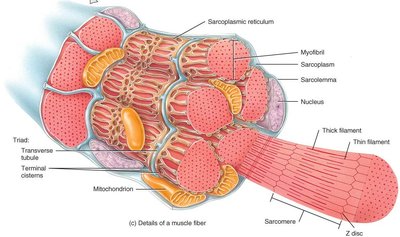
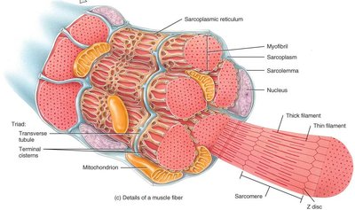
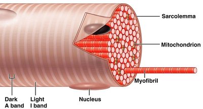
Muscle fibers (myofibers) are long, cylindrical, multinucleated cells. The sarcolemma is the cell membrane, and the sarcoplasm is the cytoplasm, filled with myofibrils and myoglobin.
Sarcolemma: Muscle cell membrane.
Sarcoplasm: Cytoplasm containing myofibrils and myoglobin.
Sarcoplasmic Reticulum (SR) and Transverse Tubules
Sarcoplasmic Reticulum (SR): Tubular sacs storing Ca2+ ions; releases Ca2+ to trigger contraction.
Transverse (T) Tubules: Invaginations of sarcolemma carrying action potentials deep into the cell.
Mitochondria: Numerous, providing ATP for contraction.
Myofibrils and Myofilaments
Myofibrils are bundles of contractile protein filaments (myofilaments) arranged in parallel, filling most of the cytoplasm. There are hundreds to thousands per cell.
Actin: Thin myofilaments.
Myosin: Thick myofilaments.
Parallel arrangement gives striated appearance.
Sarcomere: Functional Unit of Muscle Contraction
Sarcomere Structure
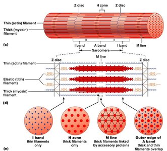
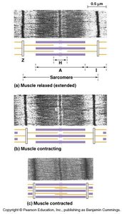
The sarcomere is the functional unit of striated muscle contraction, defined as the region between two successive Z discs. It contains organized myofilaments and shows visible banding patterns (striations).
Contains actin and myosin filaments.
Banding pattern includes A band, I band, H zone, M line, and Z disc.
Proteins in Muscle
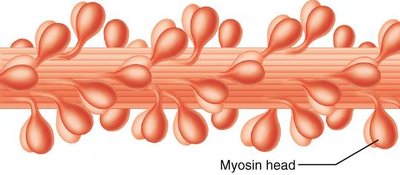
Contractile proteins: Myosin and actin.
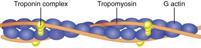
Regulatory proteins: Troponin and tropomyosin, which control contraction.
Muscle Contraction Mechanism
Role of Calcium and Regulatory Proteins
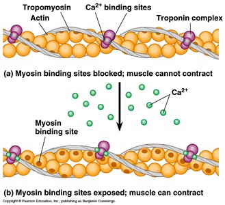
Muscle contraction is regulated by Ca2+ ions and regulatory proteins. When a muscle is relaxed, myosin binding sites on actin are blocked by tropomyosin. Upon stimulation, Ca2+ binds to troponin, causing a conformational change that exposes the binding sites, allowing contraction.
Action potential triggers Ca2+ release from SR.
Ca2+ binds to troponin, shifting tropomyosin.
Myosin binds to actin, initiating contraction.
Ca2+ pumps remove Ca2+ when stimulus ends.
Neuromuscular Junction (NMJ)
Structure and Function
The NMJ is the site of communication between a motor neuron and a muscle fiber. It consists of the synaptic cleft, synaptic vesicles containing acetylcholine (ACh), and ACh receptors on the muscle membrane.
Synapse: Small gap between neuron and muscle cell.
Synaptic cleft: Space between axon terminal and motor end plate.
Synaptic vesicles: Contain neurotransmitter (ACh).
ACh receptors: Bind ACh to initiate muscle excitation.
Destruction of Acetylcholine
Acetylcholinesterase: Enzyme that rapidly breaks down ACh in the NMJ, preventing continuous excitation.
Drugs and diseases can interfere with NMJ function (e.g., myasthenia gravis, curare).
Homeostatic Imbalances and Pharmacology
Tetanus
Caused by neurotoxin from Clostridium tetani.
Toxin blocks inhibitory nerve impulses, causing unopposed muscle contraction and spasm.
Pharmacology of the NMJ
Botulinum toxin: Blocks neurotransmitter release, causing paralysis (used in Botox).
Curare: Blocks ACh receptors, causing paralysis (used as muscle relaxant in surgery).
Neostigmine: Anticholinesterase agent, strengthens weak contractions in myasthenia gravis, antidote for curare.
Summary and Key Concepts
NMJ is critical for muscle contraction; requires nerve stimulus, ACh release, functional ACh receptor, timely Ca2+ release, actin-myosin binding, and ATP.
Sliding filament model explains striated muscle contraction.
Aging leads to replacement of skeletal muscle by fat, slowing reflexes, and decreased strength.
Exam Question
What is a muscle fiber? A) a muscle cell B) a protein C) a filament Correct answer: A) a muscle cell
