 Indietro
Muscle Tissue: Structure, Function, and Physiology
Guida di studio - Note intelligenti
 Appunti personalizzati basati sui tuoi materiali, ampliati con definizioni chiave, esempi e contesto.
Muscle Tissue
Types and Functions of Muscle Tissue
Muscle tissue is a primary tissue responsible for contraction and movement in the human body. There are three main types of muscle tissue, each with distinct functions and properties:
Skeletal muscle: Moves the body by pulling on bones.
Cardiac muscle: Pumps blood through the cardiovascular system.
Smooth muscle: Pushes fluids and solids through internal passageways and organs.
Common properties of muscle tissue include:
Excitability: Ability to receive and respond to stimuli.
Contractility: Ability of cells to shorten.
Extensibility: Ability of the muscle to stretch.
Elasticity: Ability of the muscle to recoil to its resting length.
Skeletal muscle functions include producing movement, maintaining posture, supporting soft tissues, guarding entrances and exits, maintaining body temperature, and storing nutrients.

Organization of Skeletal Muscle
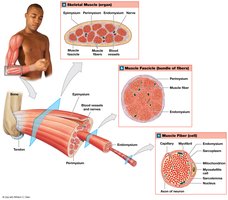
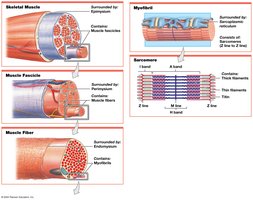
Skeletal muscles are composed of muscle tissue, connective tissues, blood vessels, and nerves. The organization at the tissue level involves three layers of connective tissue:
Epimysium: Surrounds the entire muscle and separates it from surrounding tissues.
Perimysium: Surrounds individual fascicles (bundles of muscle fibers).
Endomysium: Surrounds individual muscle cells (muscle fibers).
The collagen fibers from these layers converge to form tendons (bundles) or aponeuroses (sheets), which attach muscles to bones or other muscles.
Skeletal Muscle Fibers
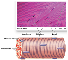
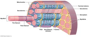
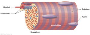
Skeletal muscle fibers are large, multinucleate, and striated due to the arrangement of myofibrils. The sarcolemma (plasma membrane) surrounds the sarcoplasm (cytoplasm) and has a resting membrane potential. Transverse tubules (T tubules) transmit action potentials deep into the cell, ensuring coordinated contraction.
Internal Structure
Sarcoplasmic reticulum (SR): Tubular network specialized for storage and release of calcium ions.
Myofibrils: Collections of myofilaments responsible for contraction.
Myofilaments: Two types—thin (actin) and thick (myosin).
Sarcomere Structure
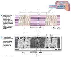
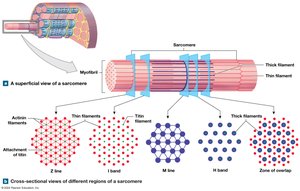
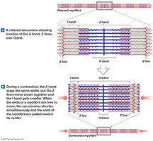
Sarcomeres are the smallest contractile units of muscle fibers, composed of organized thin and thick filaments. The arrangement of these filaments produces visible bands:
A bands: Dark bands, length of thick filaments.
M line: Center of A band, stabilizes thick filaments.
H band: Only thick filaments, either side of M line.
Zone of overlap: Region where thick and thin filaments overlap.
I bands: Light bands, only thin filaments.
Z lines: Bisect I bands, mark boundaries between sarcomeres.
Thin and Thick Filaments
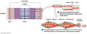
Thin filaments: Composed of F-actin (two rows of G-actin), tropomyosin (covers active sites), and troponin (binds calcium).
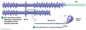
Thick filaments: Composed of myosin molecules (tail and head), with a core of titin for elasticity.
Sliding-Filament Theory
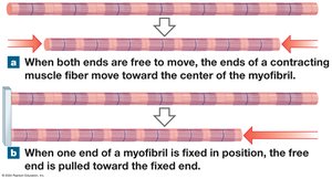
During contraction, thin filaments slide toward the M line alongside thick filaments. The H and I bands narrow, zones of overlap widen, Z lines move closer, and the A band remains constant.
Muscle Contraction
Neuromuscular Junction and Excitable Membranes
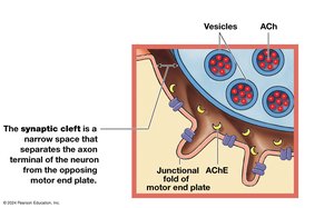
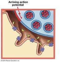
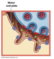
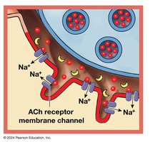
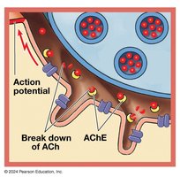
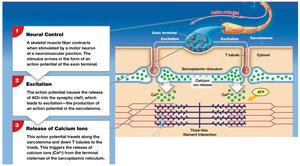
The neuromuscular junction (NMJ) is the synapse between a motor neuron and a skeletal muscle fiber. Components include:
Axon terminal: End of motor neuron.
Motor end plate: Folded membrane of muscle fiber.
Synaptic cleft: Space between axon terminal and motor end plate.
Excitation–Contraction Coupling
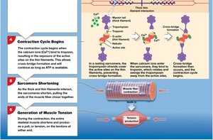
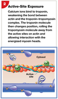
The action potential travels down T tubules to triads, triggering calcium release from the SR. Calcium binds to troponin, moving tropomyosin and exposing actin active sites, allowing contraction to begin.
Contraction Cycle and Cross-Bridge Formation
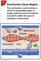
The contraction cycle involves several steps:
Arrival of calcium ions.
Exposure of active sites on actin.
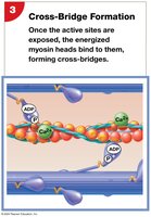
Formation of cross-bridges (myosin heads bind actin).
Power stroke (myosin head pivots, pulling actin).
Detachment and reactivation of myosin heads.
Muscle Tension and Relaxation
Muscle tension is the pull force generated during contraction. The amount of tension depends on the number of power strokes, sarcomere length, and frequency of stimulation. Relaxation occurs when ACh is broken down, calcium returns to the SR, and active sites are covered by tropomyosin.
Muscle Fiber Types and Performance
Types of Skeletal Muscle Fibers
Fast fibers: Contract quickly, large diameter, high force, fatigue quickly.
Slow fibers: Contract slowly, small diameter, high endurance, rich in mitochondria and myoglobin.
Intermediate fibers: Mid-sized, moderate endurance and force.
Muscle Hypertrophy and Atrophy
Hypertrophy: Increase in muscle size due to training.
Atrophy: Decrease in muscle size due to inactivity.
Aging and Muscle Tissue
Fibers decrease in diameter and elasticity.
Exercise tolerance and recovery decrease.
Fibrosis increases.
Muscle Fatigue and Conditioning
Anaerobic endurance: Short, intense activity supported by stored ATP and glycolysis.
Aerobic endurance: Sustained activity supported by mitochondrial ATP production.
Cross-training: Combination of aerobic and anaerobic exercises improves overall performance.
Cardiac and Smooth Muscle Tissue
Cardiac Muscle Tissue
Cardiac muscle is found only in the heart. Cells are small, branched, striated, and have a single nucleus. They are connected by intercalated discs, which allow synchronized contraction. Cardiac muscle is highly resistant to fatigue and relies on aerobic metabolism.
Smooth Muscle Tissue
Smooth muscle is found in internal organs and systems. Cells are long, slender, spindle-shaped, and nonstriated. They contract via excitation–contraction coupling involving calmodulin and myosin light chain kinase. Smooth muscle tone is maintained by neural, hormonal, or chemical factors.
Summary Table: Muscle Tissue Types
Type | Structure | Location | Function |
|---|---|---|---|
Skeletal | Striated, multinucleate | Attached to bones | Movement, posture |
Cardiac | Striated, branched, single nucleus | Heart | Pumping blood |
Smooth | Non-striated, spindle-shaped | Internal organs | Movement of materials |
Additional info: The notes expand on brief points with academic context, definitions, and examples to ensure completeness and clarity for exam preparation.
