 Indietro
Muscle Tissue: Structure, Function, and Physiology
Guida di studio - Note intelligenti
 Appunti personalizzati basati sui tuoi materiali, ampliati con definizioni chiave, esempi e contesto.
Muscle Tissue Types and Functions
Overview of Muscle Tissue
Muscle tissue is essential for movement, stability, heat generation, and the transport of substances within the body. There are three main types of muscle tissue, each with distinct structural and functional characteristics.
Skeletal muscle: Attached to bones, striated, voluntary control.
Cardiac muscle: Found in heart walls, striated, involuntary control.
Smooth muscle: Located in walls of hollow organs and blood vessels, non-striated, involuntary control.
Muscle functions include creating body movements, stabilizing positions, generating heat, storing and moving substances, and regulating blood flow.
Embryonic Development and Repair
Muscle cells originate from embryonic stem cells called myoblasts, which merge to form large, multinucleated muscle fibers. Satellite cells are stem cells reserved for minor repairs; extensive damage leads to scar tissue formation (fibrosis).
Microscopic Structure of Muscle Tissue
Striations and Sarcomeres
Striations in skeletal and cardiac muscle are visible under a light microscope and result from the arrangement of myofibrils containing repeating units called sarcomeres. Sarcomeres are the functional units of muscle contraction, defined by the space between two Z-discs.
Actin: Protein in thin filaments, pulled during contraction.
Myosin: Protein in thick filaments, uses ATP to pull actin.
Titin: Anchors Z-disc, provides elasticity.
Skeletal Muscle Structure
Skeletal muscle is organized in layers of connective tissue:
Epimysium: Surrounds entire muscle, continuous with tendon.
Perimysium: Wraps fascicles (bundles of muscle fibers).
Endomysium: Surrounds individual muscle fibers.
Fascia: Connective tissue wrapping all body parts; includes superficial and deep fascia.
Tendon: Connects muscle to bone.
Aponeurosis: Flat sheet of connective tissue for muscle attachment.
Muscle Tissue Histology
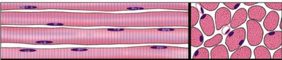
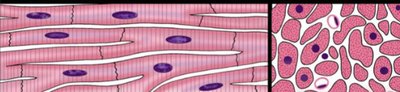
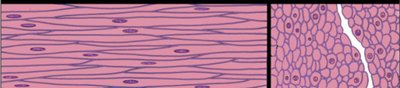
The three types of muscle tissue can be distinguished microscopically:
Skeletal muscle: Long, cylindrical, multinucleated cells with striations.
Cardiac muscle: Branched cells with striations and intercalated discs.
Smooth muscle: Spindle-shaped cells without striations.
Skeletal Muscle Proteins and Contraction
Sliding Filament Model
Muscle contraction occurs when thick and thin filaments slide past each other, shortening the sarcomere. The process is regulated by the presence of calcium ions and ATP.
Myosin heads bind to actin when binding sites are uncovered by the action of calcium binding to troponin.
ATP is required for myosin head detachment and repositioning.
Titin allows sarcomere to return to resting length after contraction.
Neuromuscular Junction (NMJ)
The NMJ is the site where a motor neuron communicates with a muscle fiber to initiate contraction.
Acetylcholine (ACh): Neurotransmitter released from synaptic vesicles.
Motor end plate: Region of muscle membrane where ACh binds.
Na+/K+ pump: Maintains ion gradients necessary for action potentials.
Acetylcholinesterase: Enzyme that breaks down ACh.
Excitation-Contraction Coupling
From NMJ to Sarcomere
Action potentials travel along the sarcolemma and T-tubules, triggering calcium release from the sarcoplasmic reticulum (SR). Calcium binds to troponin, allowing myosin to interact with actin and initiate contraction.
Terminal cisternae: SR structures storing calcium.
Calcium pump: Returns Ca++ to SR after contraction.
Excitation-contraction coupling: Link between electrical excitation and mechanical contraction.
ATP Sources in Muscle Cells
Energy Production
Muscle cells utilize several sources for ATP production:
Creatine phosphate: Rapid ATP generation, exhausted in 15 seconds.
Glycolysis: Produces 2 ATP per glucose (anaerobic).
Aerobic respiration: Produces up to 36 ATP per glucose in mitochondria.
Glycogen: Storage form of glucose in muscle.
Myoglobin: Oxygen storage in muscle.
Hemoglobin: Oxygen transport in blood.
Sarcomere Length-Tension Relationship
Optimal Tension Generation
The amount of tension generated depends on the initial sarcomere length. Maximum tension occurs when there is optimal overlap between actin and myosin, allowing numerous crossbridges to form.
Muscle Fiber Types
SO, FG, and FOG Fibers
Muscle fibers are classified based on their metabolic properties and contraction speed:
Type 1/SO (slow oxidative): Small, dark red, endurance, high myoglobin, many mitochondria.
Type 2/FG (fast glycolytic): Large, pale, strength, low myoglobin, more glycogen.
Type 2A/FOG (fast oxidative glycolytic): Intermediate, both endurance and strength.
Muscle fiber composition varies by muscle function, training, and genetics.
Motor Units and Muscle Contraction
Motor Unit Structure and Function
A motor unit consists of a motor neuron and all the muscle fibers it innervates. Motor units vary in size and are recruited based on the force required for a task.
Small motor units: Fine control (e.g., fingers).
Large motor units: Powerful movements (e.g., thighs).
Recruitment: Activation of additional motor units increases contraction strength.
Myographs and Twitch Contractions
A myogram records muscle contraction. A twitch contraction consists of latent, contraction, and relaxation periods. Fast and slow twitch fibers differ in contraction speed.
Control of Motor Units
Recruitment and Muscle Tone
The nervous system controls contraction strength and duration by varying the number and frequency of motor unit activation. Recruitment allows for smooth, sustained contractions and delays fatigue.
Muscle tone: Weak, involuntary contractions maintain posture.
Flaccid: Loss of muscle tone due to nerve damage.
Types of Muscle Contractions and Adaptations
Isotonic and Isometric Contractions
Isotonic contraction: Muscle changes length (concentric: shortens, eccentric: lengthens).
Isometric contraction: Muscle generates tension without changing length.
Atrophy, Hypertrophy, and Hyperplasia
Atrophy: Decrease in muscle size due to disuse or denervation.
Hypertrophy: Increase in muscle fiber diameter from repetitive activity.
Hyperplasia: Increase in cell number; occurs in smooth muscle, not skeletal or cardiac after birth.
Cardiac Muscle
Structure and Function
Cardiac muscle cells are connected by intercalated discs containing desmosomes and gap junctions, allowing coordinated contraction. Cells have striations, many mitochondria, and a central nucleus.
Smooth Muscle
Structure and Function
Smooth muscle lacks sarcomeres and striations. Dense bodies and intermediate filaments transmit tension. Single-unit smooth muscle cells are connected by gap junctions, while multi-unit cells require direct neural stimulation. Some organs have multiple muscle layers for complex movements.
Additional info: The included images visually reinforce the histological differences between skeletal, cardiac, and smooth muscle tissues, supporting the explanations in the corresponding paragraphs.
