Indietro
Muscle Tissue: Structure, Function, and Types
Guida di studio - Note intelligenti
 Appunti personalizzati basati sui tuoi materiali, ampliati con definizioni chiave, esempi e contesto.
Muscle Tissue
Types of Muscle Tissue
Muscle tissue is essential for movement and force generation in the human body. There are three primary types of muscle tissue, each with distinct structural and functional characteristics:
Skeletal Muscle: Voluntary, striated muscle attached to bones for movement.
Cardiac Muscle: Involuntary, striated muscle found only in the heart.
Smooth Muscle: Involuntary, non-striated muscle found in walls of internal organs.
Functions of Skeletal Muscle Tissue
Six Major Functions
Skeletal muscle tissue performs several critical functions:
Produce skeletal movement: Facilitates voluntary movement of the body.
Maintain posture and body position: Stabilizes joints and maintains posture.
Support soft tissues: Protects internal organs.
Guard entrances and exits: Controls openings of digestive and urinary tracts.
Maintain body temperature: Generates heat during contraction.
Store nutrient reserves: Stores glycogen and other nutrients.
Organization of Skeletal Muscle
Structural Hierarchy
Skeletal muscle is organized to optimize function and force transmission. The structural hierarchy includes:
Muscle tissue (muscle fibers): The contractile cells.
Connective tissues: Support and protect muscle fibers.
Nerves: Stimulate contraction.
Blood vessels: Supply nutrients and oxygen.
Three layers of connective tissue surround and organize muscle fibers:
Epimysium: Surrounds the entire muscle.
Perimysium: Surrounds bundles of muscle fibers (fascicles).
Endomysium: Surrounds individual muscle fibers.
At the ends of muscles, these connective tissues combine to form tendons (bundles) or aponeuroses (sheets).
Characteristics of Skeletal Muscle Fibers
Cellular Structure
Skeletal muscle fibers are long, multinucleate cells formed by the fusion of myoblasts. Key components include:
Sarcolemma: The cell membrane of a muscle fiber.
Sarcoplasm: The cytoplasm of a muscle fiber.
Transverse tubules (T tubules): Invaginations of the sarcolemma that transmit action potentials.
Myofibrils: Bundles of myofilaments (actin and myosin) organized into sarcomeres.
Sarcoplasmic reticulum (SR): Specialized endoplasmic reticulum for calcium storage.
Sarcomere Structure and Function
Contractile Unit of Muscle
The sarcomere is the fundamental contractile unit of muscle, composed of thick (myosin) and thin (actin) filaments. Structural features include:
A bands: Dark, thick filaments.
I bands: Light, thin filaments.
H bands: Region with only thick filaments.
M line: Center of the H band.
Z line: Boundary of the sarcomere, bisects the I band.
Sliding Filament Theory
Mechanism of Muscle Contraction
Muscle contraction occurs when thin filaments slide toward the M line, alongside thick filaments. The width of the A band remains constant, while Z lines move closer together, shortening the sarcomere.
Neuromuscular Junction and Excitation-Contraction Coupling
Sequence of Events
The neuromuscular junction (NMJ) is the site where a motor neuron stimulates a muscle fiber. The process involves:
Release of acetylcholine (ACh) from the neuron.
ACh binds to receptors on the motor end plate, opening sodium channels.
Influx of sodium ions generates an action potential in the sarcolemma.
Action potential triggers calcium release from the sarcoplasmic reticulum.
Calcium binds to troponin, exposing active sites on actin.
Myosin heads bind to actin, forming cross-bridges and initiating contraction.
The Contraction Cycle
Steps of Cross-Bridge Formation
The contraction cycle consists of several steps:
Calcium ions arrive at the zone of overlap in the sarcomere.
Active sites on actin are exposed.
Myosin heads bind to actin, forming cross-bridges.
Myosin head pivots, performing a power stroke.
Cross-bridge detachment occurs when ATP binds to myosin.
Myosin reactivation as ATP is hydrolyzed.
Tension Production and Contraction Types
Muscle Twitch and Tension
A muscle twitch consists of three phases:
Latent period: Action potential triggers calcium release.
Contraction phase: Calcium binds, tension builds.
Relaxation phase: Calcium levels fall, tension decreases.
Effects of Repeated Stimulations
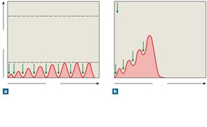
Repeated neural stimulation affects muscle tension:
Treppe: Gradual increase in tension with repeated stimuli after relaxation.
Wave summation: Increased tension when stimuli arrive before relaxation is complete.
Motor Units and Recruitment
A motor unit consists of a motor neuron and all the muscle fibers it controls. Recruitment increases tension by activating more motor units.
Muscle Tone
Muscle tone is the tension and firmness of a muscle at rest, important for maintaining posture and metabolic activity.
Types of Contraction
Isotonic contraction: Muscle changes length (concentric: shortens, eccentric: lengthens).
Isometric contraction: Muscle develops tension without changing length.
Energy for Muscle Contraction
ATP Generation
Muscle fibers use several mechanisms to generate ATP:
Creatine phosphate (CP): Stores energy for rapid ATP regeneration.
Aerobic metabolism: Occurs in mitochondria, produces 32-34 ATP per glucose.
Anaerobic glycolysis: Occurs in cytoplasm, produces 2 ATP per glucose.
Muscle Fatigue
Muscle fatigue is the reduction in power generation due to:
Fuel depletion
Accumulation of metabolic byproducts
Loss of Na+ and K+ gradients
Oxygen deficit
Types of Skeletal Muscle Fibers
Classification and Properties
Skeletal muscle fibers are classified based on contraction speed and endurance:
Fast fibers: Large diameter, rapid contraction, fatigue quickly.
Slow fibers: Small diameter, slow contraction, resistant to fatigue.
Intermediate fibers: Properties between fast and slow fibers.
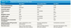
Property | Slow Fibers | Intermediate Fibers | Fast Fibers |
|---|---|---|---|
Cross-sectional diameter | Small | Intermediate | Large |
Time to peak tension | Prolonged | Medium | Rapid |
Contraction speed | Slow | Fast | Fast |
Fatigue resistance | High | Intermediate | Low |
Color | Red | Pink | White |
Myoglobin content | High | Low | Low |
Mitochondria | Many | Few | Few |
Glycolytic enzyme concentration | Low | High | High |
Substrates used for ATP generation | Lipids, carbohydrates, amino acids | Primarily carbohydrates | Carbohydrates (anaerobic) |
Alternative names | Type I, slow twitch, SO fibers | Type II-A, fast twitch, FOG fibers | Type II-B, fast twitch, FG fibers |
Muscle Growth and Atrophy
Hypertrophy and Atrophy
Hypertrophy: Increase in muscle size due to heavy training, increased myofibrils.
Atrophy: Decrease in muscle size, tone, and power due to inactivity.
Cardiac Muscle Tissue
Structure and Function
Cardiac muscle tissue is found only in the heart and is specialized for continuous rhythmic contraction. Features include:
Striated cells (cardiocytes) with a single nucleus.
Short, wide T tubules; no triads.
SR lacks terminal cisternae.
High myoglobin and mitochondria content.
Intercalated discs with gap junctions and desmosomes for mechanical, chemical, and electrical connectivity.
Cardiac muscle exhibits automaticity (pacemaker cells), variable contraction tension, extended contraction time, and prevention of tetanic contractions.
Smooth Muscle Tissue
Structure and Function
Smooth muscle tissue is found in the walls of internal organs and blood vessels. Characteristics include:
Nonstriated, spindle-shaped cells with a single central nucleus.
No T tubules, myofibrils, or sarcomeres.
Thin filaments attached to dense bodies.
Contractions transmitted from cell to cell via dense bodies.
Contraction is controlled by motor neurons or pacesetter cells.
Comparison of Muscle Tissue Types
Key Differences
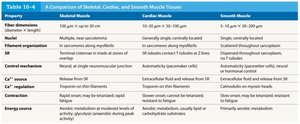
Property | Skeletal Muscle | Cardiac Muscle | Smooth Muscle |
|---|---|---|---|
Fiber dimension | Long, cylindrical | Short, branched | Spindle-shaped |
Striations | Present | Present | Absent |
SR | Extensive, terminal cisternae | Less extensive, no terminal cisternae | Scanty |
Control mechanism | Neural | Automaticity, neural | Neural, hormonal, pacesetter cells |
Ca2+ source | SR | SR and extracellular | SR and extracellular |
Contraction | Rapid, short duration | Slow, extended duration | Slow, sustained |
Energy sources | Aerobic and anaerobic | Aerobic | Primarily aerobic |