 Indietro
Muscles and Control of Body Movement: Structure, Function, and Neural Regulation
Guida di studio - Note intelligenti
 Appunti personalizzati basati sui tuoi materiali, ampliati con definizioni chiave, esempi e contesto.
Muscle Types and Their Characteristics
Overview of Muscle Types
Muscle tissue in the human body is classified into three main types, each with distinct structural and functional properties. Understanding these differences is essential for grasping how the body produces movement and maintains homeostasis.
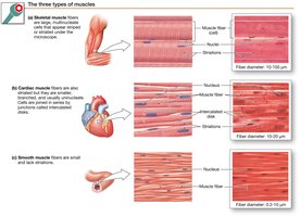
Skeletal Muscle: Striated, voluntary muscle attached to bones; responsible for body movement and posture. Controlled by somatic motor neurons.
Cardiac Muscle: Striated, involuntary muscle found only in the heart; contracts spontaneously and is regulated by the autonomic nervous system and hormones.
Smooth Muscle: Non-striated, involuntary muscle found in walls of internal organs and blood vessels; contracts slowly and can sustain contractions for long periods.
Skeletal Muscle Structure
Organization of Skeletal Muscle
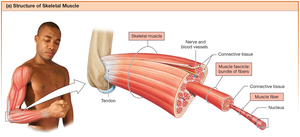
Skeletal muscle is composed of bundles of muscle fibers (cells), which are grouped into fascicles and surrounded by connective tissue. This organization allows for efficient force transmission and attachment to bones via tendons.
Muscle Fiber: The basic cellular unit of muscle, also called a muscle cell.
Fascicle: A bundle of muscle fibers surrounded by connective tissue.
Connective Tissue: Surrounds individual fibers, fascicles, and the entire muscle, merging with tendons to attach muscle to bone.
Muscle Fiber Anatomy and Sarcomere Structure
Specialized Structures in Muscle Fibers
Muscle fibers contain specialized organelles and structures that facilitate contraction:
Sarcolemma: The plasma membrane of a muscle fiber.
Sarcoplasm: The cytoplasm of a muscle fiber.
Sarcoplasmic Reticulum (SR): Specialized endoplasmic reticulum that stores calcium ions (Ca2+).
Transverse Tubules (T-tubules): Invaginations of the sarcolemma that transmit action potentials into the fiber's interior.
Myofibrils: Bundles of contractile proteins (myofilaments) organized into repeating units called sarcomeres.
Sarcomere: The Functional Unit of Contraction
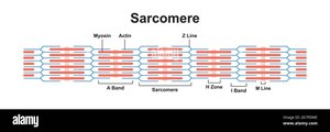
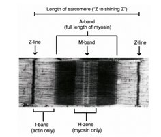
The sarcomere is the basic contractile unit of muscle, defined as the region between two Z disks. Its precise arrangement of thick and thin filaments gives skeletal and cardiac muscle their striated appearance.
Z disk: Anchors thin filaments (actin); marks the boundary of each sarcomere.
I band: Contains only thin filaments (actin).
A band: Length of thick filaments (myosin); includes regions of overlap with actin.
H zone: Central region of A band with only thick filaments.
M line: Center of the sarcomere; holds thick filaments together.
Myofilaments: Actin and Myosin
Myofibrils are composed of two main types of protein filaments:
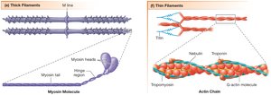
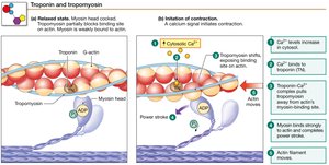
Thin Filaments (Actin): Composed of F-actin (filamentous actin), tropomyosin, and troponin. Tropomyosin blocks myosin-binding sites on actin; troponin binds Ca2+ to initiate contraction.
Thick Filaments (Myosin): Made of myosin molecules with heads that bind to actin and hydrolyze ATP for energy.
Titin: An elastic protein that stabilizes myosin and provides elasticity to the sarcomere.
Nebulin: An inelastic protein that helps align actin filaments.
Mechanism of Muscle Contraction
Sliding Filament Theory
Muscle contraction occurs when thin filaments slide past thick filaments, shortening the sarcomere without changing the length of the filaments themselves. This process is powered by ATP hydrolysis and regulated by Ca2+ binding to troponin.
Sequence of Events in Muscle Contraction
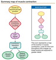
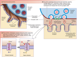
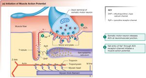
Events at the Neuromuscular Junction (NMJ): Action potential in a motor neuron triggers release of acetylcholine (ACh), which binds to nicotinic receptors on the muscle fiber, causing depolarization.
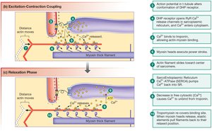
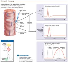
Excitation-Contraction (E-C) Coupling: The action potential travels along the sarcolemma and T-tubules, triggering Ca2+ release from the SR.
Contraction-Relaxation Cycle: Ca2+ binds to troponin, allowing myosin to bind actin and initiate the crossbridge cycle. Relaxation occurs when Ca2+ is pumped back into the SR.
Neuromuscular Junction and Action Potential Generation
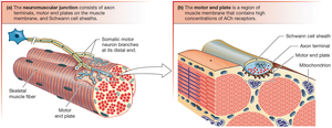
The NMJ is a specialized synapse where a motor neuron communicates with a skeletal muscle fiber. The arrival of an action potential at the axon terminal causes ACh release, which opens ligand-gated ion channels on the muscle membrane, leading to depolarization and initiation of a muscle action potential.
Excitation-Contraction Coupling and Calcium Release
Depolarization of the T-tubules activates voltage-gated DHP receptors, which mechanically open RyR Ca2+ channels in the SR, releasing Ca2+ into the sarcoplasm. This Ca2+ binds to troponin, shifting tropomyosin and exposing myosin-binding sites on actin.
Crossbridge Cycle
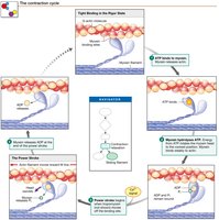
The crossbridge cycle describes the sequence of events by which myosin heads bind to actin, perform a power stroke, and detach, powered by ATP hydrolysis:
ATP binds to myosin, causing it to detach from actin.
ATP is hydrolyzed, energizing the myosin head.
Myosin binds to actin, forming a crossbridge.
Release of Pi initiates the power stroke, pulling actin toward the M line.
ADP is released, and the cycle repeats as long as Ca2+ and ATP are present.
Muscle Relaxation
Relaxation occurs when Ca2+ is actively transported back into the SR by Ca2+-ATPase pumps. As Ca2+ levels in the sarcoplasm fall, troponin and tropomyosin return to their resting positions, blocking myosin-binding sites on actin and ending contraction.
Muscle Twitch and Summation
Muscle Twitch and Phases
A muscle twitch is a single contraction-relaxation cycle in response to a single stimulus. It consists of three phases:
Latent Period: Time between stimulus and onset of contraction; includes E-C coupling and Ca2+ release.
Contraction Phase: Muscle tension rises as crossbridges form.
Relaxation Phase: Ca2+ is reabsorbed, and tension falls.
Summation and Tetanus
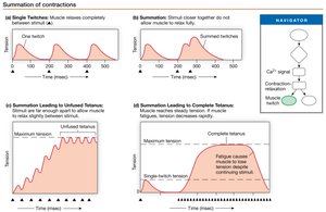
When stimuli are delivered in rapid succession, muscle fibers do not fully relax between contractions, resulting in summation of tension. If stimulation is frequent enough, a sustained maximal contraction called tetanus occurs.
Motor Units and Force Generation
Motor Units
A motor unit consists of a single somatic motor neuron and all the muscle fibers it innervates. The force of muscle contraction depends on the number and type of motor units recruited.
Recruitment: Increasing the number of active motor units increases the overall force generated by the muscle.
Length-Tension Relationship
The amount of tension a muscle can generate depends on the initial length of its sarcomeres. Maximum force is produced when sarcomeres are at an optimal length, allowing the greatest number of crossbridges to form.
Muscle Metabolism and Fatigue
ATP Sources for Muscle Contraction
Skeletal muscle requires a continuous supply of ATP for contraction, which is generated by three main pathways:
Aerobic Metabolism: Uses glucose and oxygen in the citric acid cycle and electron transport chain; efficient but slower.
Anaerobic Glycolysis: Produces ATP quickly but less efficiently; results in lactate formation.
Phosphocreatine Breakdown: Provides a rapid, short-term supply of ATP via the enzyme creatine kinase.
Muscle Fatigue
Fatigue is a decline in muscle tension due to prolonged activity. It can result from central (CNS) or peripheral (muscle or neuron) factors, including depletion of energy stores, accumulation of metabolic byproducts, and ion imbalances.
Types of Skeletal Muscle Fibers
Classification by Speed and Fatigue Resistance
Skeletal muscle fibers are classified based on their contraction speed and resistance to fatigue:
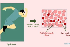
Slow-Twitch (Type I): Rely on oxidative phosphorylation; fatigue-resistant; high myoglobin content.
Fast-Twitch Oxidative-Glycolytic (Type IIa): Use both aerobic and anaerobic metabolism; intermediate fatigue resistance.
Fast-Twitch Glycolytic (Type IIb/X): Rely primarily on anaerobic glycolysis; fatigue quickly; low myoglobin content.
Mechanics of Body Movement
Types of Muscle Contractions
Muscle contractions are classified based on whether they produce movement:
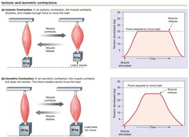
Isotonic Contractions: Muscle changes length and moves a load. Includes concentric (shortening) and eccentric (lengthening) actions.
Isometric Contractions: Muscle generates force without changing length; elastic elements stretch to absorb tension.
Smooth Muscle Structure and Function
Organization and Properties
Smooth muscle cells are spindle-shaped, have a single central nucleus, and lack striations. They contain dense bodies (analogous to Z disks) and intermediate filaments for structural support. Smooth muscle contracts and relaxes more slowly than skeletal muscle and can sustain contractions for extended periods without fatigue.
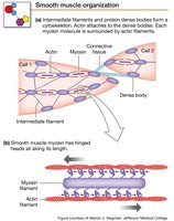
Types of Smooth Muscle Contractions
Smooth muscle can exhibit different contraction patterns, including tonic (sustained) and phasic (rhythmic) contractions, depending on the organ and physiological need.
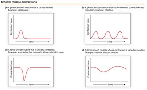
Coordination of Smooth Muscle Activity
Smooth muscle cells communicate via gap junctions in single-unit (visceral) smooth muscle, allowing coordinated contraction. In multiunit smooth muscle, cells contract independently, allowing finer control (e.g., in the eye).
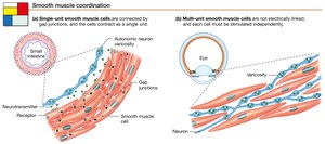
Smooth Muscle Contraction and Relaxation Mechanisms
Smooth muscle contraction can be triggered by electrical or chemical signals. Ca2+ influx leads to phosphorylation of myosin light chains, activating myosin ATPase and initiating contraction. Relaxation occurs when myosin light chain phosphatase (MLCP) dephosphorylates myosin, sometimes resulting in a latch state where myosin remains attached to actin.
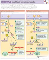
Cardiac Muscle Structure and Function
Cardiac Muscle Properties
Cardiac muscle shares features with both skeletal and smooth muscle. It is striated and organized into sarcomeres but consists of shorter, branched fibers with a single nucleus. Cardiac muscle cells are electrically connected via gap junctions in intercalated disks and can generate pacemaker potentials. They are regulated by both the autonomic nervous system and hormones.
Neural Control of Muscle Contraction
Neural Reflexes
Neural reflexes are automatic responses to stimuli and can be classified by their efferent division (somatic or autonomic), integration site (spinal or cranial), developmental origin (innate or learned), and number of synapses (monosynaptic or polysynaptic).
Monosynaptic Reflex: Stretch Reflex
The stretch reflex (e.g., patellar tendon reflex) is a monosynaptic reflex involving muscle spindles that monitor muscle length. Alpha motor neurons stimulate extrafusal fibers, while gamma motor neurons adjust intrafusal fiber sensitivity.
Disynaptic Reflex: Golgi Tendon Organ
Golgi tendon organs detect muscle tension and mediate inhibitory reflexes to prevent excessive force generation, protecting muscles and tendons from injury.
Polysynaptic Reflex: Reciprocal Inhibition and Crossed Extensor Reflex
Reciprocal inhibition ensures that antagonistic muscles relax as prime movers contract. The crossed extensor reflex coordinates withdrawal of a limb from a painful stimulus while extending the opposite limb for balance.
Autonomic (Visceral) Reflexes
Autonomic reflexes control smooth and cardiac muscle activity and can be modulated by the brain. Some fibers in these tissues can spontaneously depolarize, acting as pacemakers.
Summary Table: Muscle Types Comparison
Feature | Skeletal Muscle | Cardiac Muscle | Smooth Muscle |
|---|---|---|---|
Striations | Yes | Yes | No |
Control | Voluntary | Involuntary | Involuntary |
Location | Skeletal system | Heart | Internal organs, vessels |
Cell Shape | Long, cylindrical, multinucleate | Short, branched, single nucleus | Spindle-shaped, single nucleus |
Pacemaker Activity | No | Yes (some cells) | Yes (some cells) |
Regeneration | Limited | Limited | Good |
Key Questions for Review
What is a sarcomere and its components?
What is the neuromuscular junction? How are action potentials generated in the muscle?
What are the steps in the excitation-contraction coupling mechanism?
What are the types of muscle responses and their characteristics?
How does skeletal muscle obtain its energy?
Describe the types of skeletal muscle fibers.
How does the nervous system control contraction?
How is smooth muscle contraction different from skeletal muscle?
