 Indietro
Muscles and Muscle Tissue: Structure, Function, and Contraction
Guida di studio - Note intelligenti
 Appunti personalizzati basati sui tuoi materiali, ampliati con definizioni chiave, esempi e contesto.
Muscle Tissue Overview
Types of Muscle Tissue
Muscle tissue is essential for movement and comprises nearly half of the body's mass. It transforms chemical energy (ATP) into mechanical energy, enabling force generation. There are three primary types of muscle tissue, each with distinct characteristics and functions:
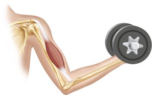
Skeletal Muscle: Attached to bones and skin, responsible for voluntary movements. Fibers are long, striated, and can be consciously controlled. They contract rapidly but tire easily.
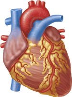
Cardiac Muscle: Found only in the heart, responsible for pumping blood. Fibers are striated but involuntary, controlled by the heart's pacemaker and modulated by the nervous system.
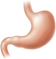
Smooth Muscle: Located in walls of hollow organs (e.g., stomach, bladder, airways). Fibers are non-striated and involuntary, controlling movements like digestion and airway regulation.
Muscle Type | Location | Striations | Control |
|---|---|---|---|
Skeletal | Bones, skin | Striated | Voluntary |
Cardiac | Heart | Striated | Involuntary |
Smooth | Hollow organs | Non-striated | Involuntary |
Characteristics of Muscle Tissue
All muscle tissues share four fundamental properties:
Excitability: Ability to respond to stimuli.
Contractility: Ability to shorten forcibly when stimulated.
Extensibility: Ability to be stretched.
Elasticity: Ability to recoil to resting length after stretching.
Functions of Muscle Tissue
Produce Movement: Enables locomotion and manipulation.
Maintain Posture: Stabilizes body position.
Stabilize Joints: Supports joint integrity.
Generate Heat: Maintains body temperature during contraction.
Skeletal Muscle Structure
Organizational Levels
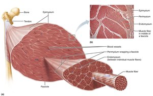
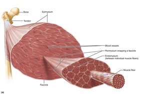
Skeletal muscle is an organ composed of muscle fibers, nerves, blood vessels, and connective tissue sheaths. The connective tissue sheaths provide structural support and organization:
Epimysium: Surrounds the entire muscle.
Perimysium: Surrounds groups of muscle fibers (fascicles).
Endomysium: Surrounds individual muscle fibers.
Muscle Attachments
Direct Attachment: Epimysium fused to periosteum of bone or perichondrium of cartilage.
Indirect Attachment: Connective tissue extends beyond muscle as a tendon or aponeurosis.
Skeletal Muscle Fibers
Microscopic Anatomy
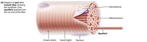
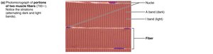
Skeletal muscle fibers are long, cylindrical cells with multiple nuclei. Key structures include:
Sarcolemma: Plasma membrane of the muscle fiber.
Sarcoplasm: Cytoplasm containing glycosomes (glycogen storage) and myoglobin (oxygen storage).
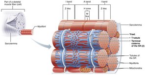
Myofibrils: Densely packed rodlike elements responsible for striations and contraction.
Sarcoplasmic Reticulum: Specialized endoplasmic reticulum for calcium storage and release.
T Tubules: Extensions of the sarcolemma that transmit electrical signals deep into the fiber.
Myofibril Structure
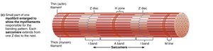
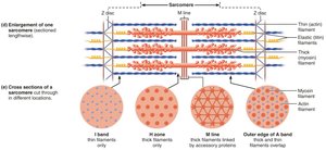
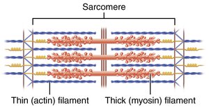
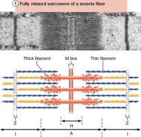
Striations: Alternating dark (A bands) and light (I bands) regions.
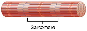
Sarcomere: The functional contractile unit, defined as the region between two Z discs.
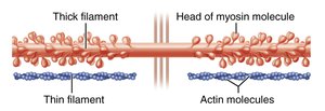
Myofilaments: Actin (thin) and myosin (thick) filaments arranged in a hexagonal pattern.
Molecular Composition of Myofilaments
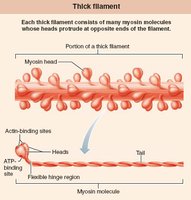
Thick Filaments: Composed of myosin molecules with globular heads that form cross bridges during contraction.
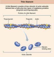
Thin Filaments: Composed of actin, tropomyosin, and troponin. Actin subunits provide binding sites for myosin heads.
Elastic Filament: Made of titin, helps maintain sarcomere structure and elasticity.
Dystrophin: Links thin filaments to sarcolemma proteins.
Clinical Application: Duchenne Muscular Dystrophy (DMD)
Pathophysiology
DMD is a severe, sex-linked recessive muscular dystrophy affecting mostly males. It is caused by a defective gene for dystrophin, leading to fragile sarcolemma, excess calcium entry, muscle fiber damage, inflammation, and loss of regenerative capacity.
Symptoms: Clumsiness, frequent falls, progressive muscle weakness.
Progression: Starts in extremities, advances to head, chest, and cardiac muscles.
Treatment: No cure; corticosteroids and supportive care can prolong life. Gene therapy is emerging.

Sarcoplasmic Reticulum and T Tubules
Structure and Function
Sarcoplasmic Reticulum (SR): Network surrounding each myofibril, stores and releases calcium ions.
T Tubules: Invaginations of the sarcolemma, allow electrical signals to penetrate deep into the muscle fiber.
Triad: Consists of a T tubule flanked by two terminal cisterns of the SR, crucial for excitation-contraction coupling.
Sliding Filament Model of Contraction
Mechanism of Muscle Contraction
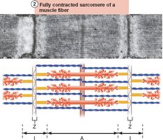
Muscle contraction occurs when myosin heads bind to actin, forming cross bridges. The sliding filament model explains how thin filaments slide past thick filaments, increasing overlap and shortening the sarcomere without changing filament length.
Key Events: Cross bridge formation, ratcheting action, shortening of I bands, disappearance of H zones, and approximation of Z discs.
Motor Neurons and Muscle Contraction
Neuromuscular Junction and Action Potential
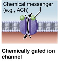
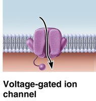
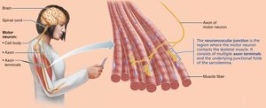
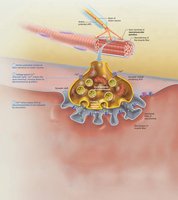
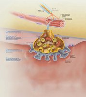
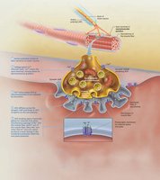
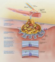
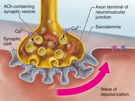
Motor neurons stimulate skeletal muscle fibers via the neuromuscular junction. The process involves transmission of an action potential, release of acetylcholine (ACh), and activation of ion channels.
Ion Channels:
Chemically gated (opened by neurotransmitters like ACh)
Voltage-gated (opened by changes in membrane potential)
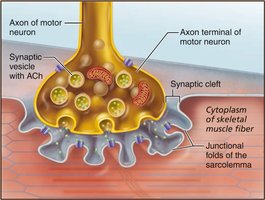
Neuromuscular Junction Anatomy
Axon Terminal: Contains synaptic vesicles with ACh.
Synaptic Cleft: Gel-filled space separating axon terminal and muscle fiber.
Junctional Folds: Sarcolemma infoldings with ACh receptors.
Events at the Neuromuscular Junction
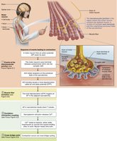
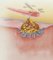
Action potential arrives at axon terminal.
Voltage-gated calcium channels open; calcium enters motor neuron.
Calcium triggers release of ACh into synaptic cleft.
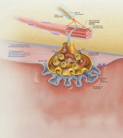
ACh binds to receptors, opening ion channels and generating end plate potential.
Acetylcholinesterase degrades ACh, terminating the signal.
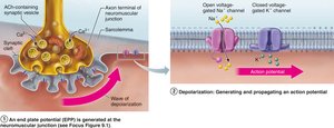
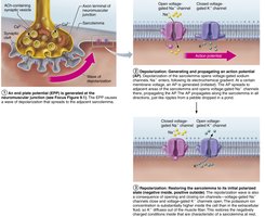
Action Potential Generation and Propagation
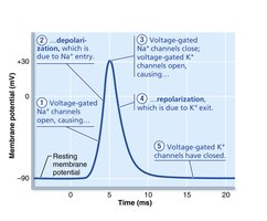
Phases of Action Potential
Resting State: Sarcolemma is polarized; inside is negative relative to outside.
Depolarization: Influx of sodium ions (Na+) makes inside more positive.
Repolarization: Efflux of potassium ions (K+) restores negative membrane potential.
Refractory Period: Fiber cannot be stimulated until repolarization is complete.
Excitation-Contraction Coupling
Linking Electrical and Mechanical Events
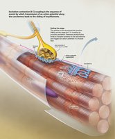
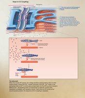
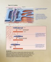
Excitation-contraction (E-C) coupling describes how an action potential leads to muscle contraction. The action potential travels along the sarcolemma and into T tubules, triggering calcium release from the SR, which initiates the sliding of myofilaments.
Key Steps:
Action potential propagates along sarcolemma and T tubules.
Voltage-sensitive proteins stimulate calcium release from SR.
Calcium binds to troponin, exposing actin binding sites.
Cross bridge cycling and contraction occur.
Summary Table: Comparison of Muscle Types
Feature | Skeletal | Cardiac | Smooth |
|---|---|---|---|
Location | Bones, skin | Heart | Hollow organs |
Striations | Yes | Yes | No |
Control | Voluntary | Involuntary | Involuntary |
Cell Shape | Long, cylindrical | Branched | Spindle-shaped |
Nuclei | Multinucleate | 1-2 per cell | Single |
Key Equations
Resting Membrane Potential
The resting membrane potential is determined by ion concentrations and permeability:
Sliding Filament Model
Muscle force generation is proportional to the number of cross bridges formed:
Clinical Note: Myasthenia Gravis
Pathophysiology
Myasthenia gravis is an autoimmune disease where antibodies destroy ACh receptors, leading to muscle weakness, drooping eyelids, and difficulty swallowing.
Treatment: Immunosuppressants and acetylcholinesterase inhibitors.
Conclusion
Understanding muscle tissue structure, function, and contraction mechanisms is fundamental for diagnosing and treating muscular disorders, and for appreciating the complexity of human movement and physiology.
