 Indietro
Muscles & Muscle Tissue: Structure, Function, and Physiology
Guida di studio - Note intelligenti
 Appunti personalizzati basati sui tuoi materiali, ampliati con definizioni chiave, esempi e contesto.
Muscle Tissue Overview
Introduction to Muscle Tissue
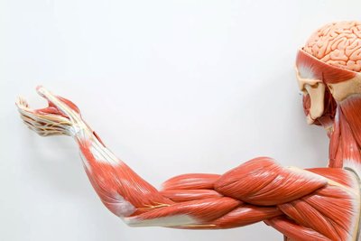
Muscle tissue is a specialized tissue responsible for movement, posture, and various physiological functions. It is classified into three types: skeletal, cardiac, and smooth muscle, each with unique structural and functional characteristics.
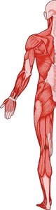
Skeletal muscle: Voluntary, striated muscle attached to bones for movement.
Cardiac muscle: Involuntary, striated muscle found in the heart.
Smooth muscle: Involuntary, non-striated muscle found in walls of hollow organs.
Functions of Skeletal Muscle
Skeletal muscle performs several essential functions in the human body:
Produce movement: Contracts to move bones and body structures.
Maintain posture and stabilize joints: Supports body position and joint stability.
Protect and support: Shields internal organs, especially in the abdominopelvic cavity.
Regulate passage of materials: Sphincters control voluntary passage of substances (e.g., urine, feces).
Produce heat: Muscle activity generates heat during contraction.
Characteristics of Muscle Tissue
Muscle tissue possesses unique properties that enable its function:
Excitability: Responds to stimuli by changing membrane potential.
Conductivity: Propagates electrical signals (action potentials).
Contractility: Generates force via actin-myosin interactions.
Extensibility: Can be stretched without damage.
Elasticity: Returns to original length after stretching.
Anatomy of Skeletal Muscle
Development of Skeletal Muscle
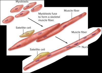
Skeletal muscle fibers are formed by the fusion of myoblasts during development. Satellite cells are muscle stem cells involved in repair and limited regeneration.
Skeletal muscle fibers: Long, multinucleated cells.
Satellite cells: Aid in muscle repair.
Sarcoplasm: Cytoplasm of muscle fibers, rich in mitochondria.
Microscopic Anatomy of Skeletal Muscle Fiber
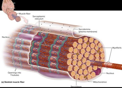
Skeletal muscle fibers contain myofibrils, which are composed of repeating units called sarcomeres. The sarcolemma is the plasma membrane, and the sarcoplasmic reticulum stores calcium ions.
Myofibrils: Bundles of myofilaments (actin and myosin).
Sarcolemma: Plasma membrane of muscle fiber.
Sarcoplasmic reticulum: Specialized endoplasmic reticulum for Ca2+ storage.
Mitochondria: Abundant for ATP production.
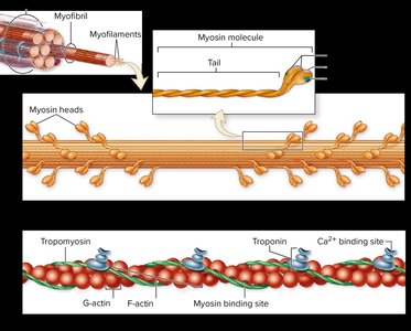
Molecular Structure of Thick & Thin Filaments
Myofibrils are composed of thick and thin filaments:
Thick filaments: Made of myosin; myosin heads bind actin and use ATP for force generation.
Thin filaments: Made of actin, tropomyosin, and troponin.
Tropomyosin: Covers myosin-binding sites on actin at rest.
Troponin: Binds Ca2+, causing tropomyosin to shift and expose binding sites.
Structure of a Sarcomere
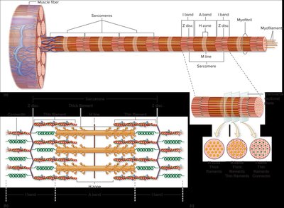
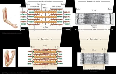
The sarcomere is the functional unit of skeletal muscle, consisting of repeating arrangements of thick and thin filaments. During contraction, thin filaments slide past thick filaments, shortening the sarcomere.
Z disc: Boundary of the sarcomere.
I band: Region with only thin filaments.
H zone: Region with only thick filaments.
A band: Region with thick filaments; remains unchanged during contraction.
Physiology of Skeletal Muscle Contraction
Motor Units and Neuromuscular Junction (NMJ)
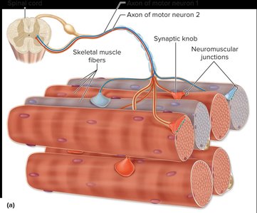
A motor unit consists of a somatic motor neuron and all the skeletal muscle fibers it innervates. The neuromuscular junction is the site where the motor neuron communicates with the muscle fiber to stimulate contraction.
Motor unit: One neuron and all its innervated muscle fibers.
NMJ: Specialized synapse for muscle activation.
Overview of Events in Skeletal Muscle Contraction
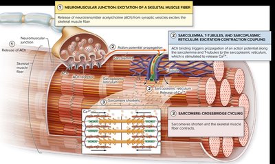
Muscle contraction involves three main steps:
Neuromuscular junction: Release of acetylcholine (ACh) triggers muscle fiber excitation.
Excitation-contraction coupling: Action potential propagates along sarcolemma and T-tubules, leading to Ca2+ release.
Crossbridge cycling: Myosin heads bind actin, causing sarcomere shortening.
Events of an Action Potential at the Sarcolemma
The sarcolemma undergoes changes in membrane potential during muscle activation:
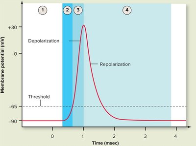
Resting membrane potential (RMP): -90 mV in unstimulated muscle fiber.
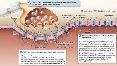
Threshold: ACh receptors open, Na+ enters, RMP changes to -65 mV.
Depolarization: Voltage-gated Na+ channels open, Na+ enters, polarity reverses to +30 mV.
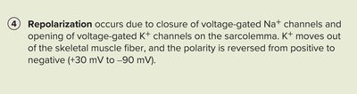
Repolarization: Na+ channels close, K+ channels open, K+ exits, polarity returns to -90 mV.



Excitation-Contraction Coupling
Excitation-contraction coupling links the action potential to muscle contraction:
ACh binds to receptors, generating end plate potential.
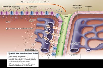
Action potential propagates along sarcolemma and T-tubules.
Ca2+ released from sarcoplasmic reticulum initiates contraction.
Crossbridge Cycling
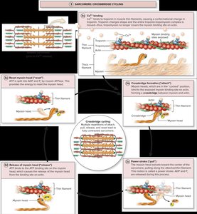
Crossbridge cycling is the process by which myosin heads bind to actin, pull, detach, and re-cock, resulting in sarcomere shortening.
Crossbridge formation: Myosin head binds to actin.
Power stroke: Myosin head pivots, pulling thin filament.
Detachment: ATP binds to myosin, causing release from actin.
Reactivation: ATP hydrolysis re-energizes myosin head.
Sarcomere Shortening
During contraction, sarcomeres shorten as thin filaments slide past thick filaments. Z discs move closer, I band and H zone decrease, while A band remains unchanged.
Muscle Tension and Metabolism
Muscle Tension in Skeletal Muscle
Muscle tension is the force produced by muscle. It can be modulated by twitch, motor unit recruitment, wave summation, and tetany.
Muscle twitch: Single, brief contraction-relaxation response.
Motor unit recruitment: Activation of additional motor units increases tension.
Wave summation: Increased tension with repeated stimulation.
Incomplete tetany: Sustained contraction with partial relaxation.
Complete tetany: Smooth, sustained contraction without relaxation.
Muscle fatigue: Decline in tension during prolonged activity.
Skeletal Muscle Metabolism
Muscle fibers generate ATP through various metabolic processes. Oxygen debt refers to the extra oxygen required after exercise to restore reserves, replenish ATP, process lactate, and return to resting conditions.
Restore oxygen reserves: Replenish myoglobin-bound oxygen.
Replenish ATP and creatine phosphate: Restore immediate energy supplies.
Process accumulated lactate: Convert lactate to fuels or glucose.
Return to resting conditions: Recover temperature, heart rate, and ventilation.
Skeletal Muscle Fiber Types
Classification of Myofiber Types
Muscle fibers are classified by contractile and metabolic characteristics:
Slow Oxidative (SO, Type I): Slow contraction, high fatigue resistance, aerobic metabolism. Example: posture, marathon running.
Fast Oxidative (FO, Type IIa): Fast contraction, moderate fatigue resistance, aerobic and glycolytic metabolism. Example: walking, cycling.
Fast Glycolytic (FG, Type IIx): Fast contraction, low fatigue resistance, glycolytic metabolism. Example: sprinting, heavy lifting.
Feature | SO (Type I) | FO (Type IIa) | FG (Type IIx) |
|---|---|---|---|
Contraction speed | Slow | Fast | Fast |
Relative force | Low | Moderate | High |
ATP production | Aerobic | Aerobic & Glycolytic | Glycolytic |
Fatigue resistance | High | Moderate | Low |
Example | Posture, marathon | Walking, cycling | Sprinting, lifting |
Cardiac Muscle Tissue
Microscopic Anatomy of Cardiac Muscle
Cardiac muscle is found in the heart wall (myocardium). Cells are short, branched, and striated, with intercalated discs containing desmosomes and gap junctions for mechanical and electrical connectivity. ATP production is primarily aerobic.
Location: Myocardium
Cell shape: Short, branched
Striations: Present
Intercalated discs: Desmosomes and gap junctions
ATP production: Aerobic, abundant mitochondria
Smooth Muscle Tissue
Microscopic Anatomy of Smooth Muscle
Smooth muscle is found in the walls of hollow organs and regulates movement and diameter. Cells are fusiform, non-striated, with a single nucleus. Contraction is regulated by calmodulin, not troponin.
Cell shape: Fusiform (spindle-shaped)
Nucleus: Single, central
Striations: Absent
T-tubules: Absent; caveolae present
Ca2+ source: Extracellular and sarcoplasmic reticulum
Regulatory protein: Calmodulin
Smooth Muscle Contraction
Unlike skeletal muscle, Ca2+ binds to calmodulin to initiate contraction. Relaxation occurs as myosin light-chain phosphatase removes phosphate groups from myosin, reducing crossbridge cycling.
Ca2+ binding: Calmodulin (not troponin)
Relaxation: MLCP removes phosphate from myosin
