 Indietro
Muscles and Muscle Tissue: Structure, Function, and Contraction
Guida di studio - Note intelligenti
 Appunti personalizzati basati sui tuoi materiali, ampliati con definizioni chiave, esempi e contesto.
Muscle Tissue Overview
Types and Characteristics of Muscle Tissue
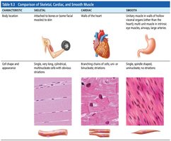
Muscle tissue is a fundamental component of the human body, accounting for nearly half of its mass. It is specialized to transform chemical energy (ATP) into mechanical energy, enabling force generation and movement. There are three primary types of muscle tissue, each with distinct structural and functional characteristics:
Skeletal Muscle: Attached to bones and skin, responsible for voluntary movements. Fibers are long, cylindrical, striated, and multinucleated.
Cardiac Muscle: Found only in the heart, responsible for pumping blood. Fibers are striated, branched, and involuntary.
Smooth Muscle: Located in walls of hollow organs (e.g., stomach, bladder, airways). Fibers are spindle-shaped, non-striated, and involuntary.
Prefixes such as myo-, mys-, and sarco- are commonly used in muscle terminology (e.g., sarcoplasm refers to muscle cell cytoplasm).
Functions of Muscle Tissue
Muscle tissue performs four essential functions:
Produce Movement: Facilitates locomotion and manipulation (e.g., walking, digestion, blood circulation).
Maintain Posture: Stabilizes body position.
Stabilize Joints: Reinforces and supports joints.
Generate Heat: Contraction produces heat, aiding in temperature regulation.
Structure and Organization of Skeletal Muscle
Nerve and Blood Supply
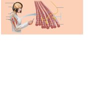
Each skeletal muscle receives a dedicated nerve, artery, and veins. Nerves supply every muscle fiber, enabling precise control. Muscles require substantial oxygen and nutrients, and waste removal is essential for function.
Connective Tissue Sheaths
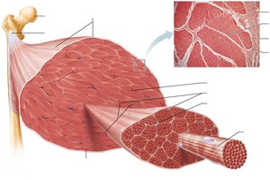
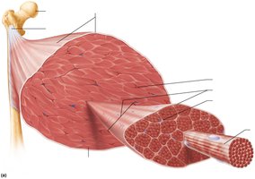
Skeletal muscle and its fibers are enveloped by connective tissue sheaths, which support and reinforce the muscle:
Epimysium: Dense irregular connective tissue surrounding the entire muscle; may blend with fascia.
Perimysium: Fibrous connective tissue surrounding fascicles (bundles of muscle fibers).
Endomysium: Fine areolar connective tissue surrounding each muscle fiber.
Muscle Attachments
Muscles span joints and attach to bones at two points:
Origin: Attachment to immovable or less movable bone.
Insertion: Attachment to movable bone.
Attachments can be direct (epimysium fused to periosteum) or indirect (connective tissue extends as tendon or aponeurosis).
Structure and Organizational Levels
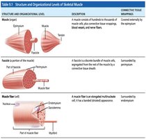
Skeletal muscle is organized hierarchically:
Muscle (organ): Composed of hundreds to thousands of muscle cells, connective tissue, blood vessels, and nerves.
Fascicle: Bundle of muscle fibers within the muscle.
Muscle Fiber (cell): Elongated multinucleate cell, surrounded by endomysium.
Muscle Fiber Microanatomy and Sliding Filament Model
Microscopic Anatomy of Skeletal Muscle Fiber
Skeletal muscle fibers are long, cylindrical cells with multiple nuclei. Key components include:
Sarcolemma: Plasma membrane of the muscle fiber.
Sarcoplasm: Cytoplasm containing glycosomes (glycogen storage) and myoglobin (O2 storage).
Myofibrils: Rod-like structures responsible for contraction.
Sarcoplasmic Reticulum (SR): Specialized endoplasmic reticulum for Ca2+ storage and release.
T Tubules: Extensions of the sarcolemma that transmit electrical signals.
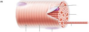
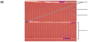
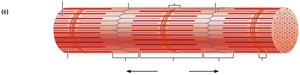
Myofibrils and Sarcomere Structure
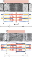
Myofibrils exhibit striations due to alternating dark (A bands) and light (I bands) regions. The sarcomere is the smallest contractile unit, defined as the segment between two Z discs. Key features include:
A Band: Dark region containing thick filaments (myosin).
I Band: Light region containing thin filaments (actin).
H Zone: Lighter region in the middle of the A band.
M Line: Protein line bisecting the H zone.
Z Disc: Coin-shaped sheet of proteins anchoring thin filaments.
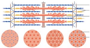
Myofilaments: Thick and Thin Filaments
Myofilaments are organized within the sarcomere:
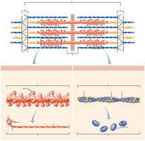
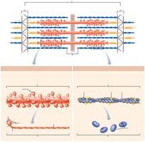
Thick Filaments: Composed of myosin molecules, each with a tail and globular head. Myosin heads form cross bridges with actin during contraction.
Thin Filaments: Composed of actin, tropomyosin, and troponin. Actin subunits provide binding sites for myosin.
Elastic Filament: Made of titin, maintains alignment and elasticity.
Sarcoplasmic Reticulum and T Tubules
Structure and Function
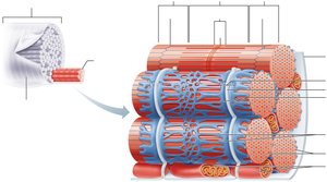
The sarcoplasmic reticulum (SR) is a network of smooth endoplasmic reticulum surrounding each myofibril, functioning in Ca2+ regulation. T tubules are invaginations of the sarcolemma that allow electrical signals to penetrate deep into the muscle fiber. The triad consists of a T tubule flanked by two terminal cisterns of the SR.
Sliding Filament Model of Contraction
Mechanism of Muscle Contraction
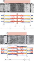
Contraction is initiated when cross bridges form between myosin heads and actin. The sliding filament model explains how thin filaments slide past thick filaments, increasing overlap and shortening the sarcomere. Neither filament changes length; instead, their relative positions shift.
In relaxed muscle, filaments overlap slightly.
Upon stimulation, myosin heads bind actin, pulling thin filaments toward the center (M line).
Repeated cross bridge cycling shortens the muscle fiber.
Muscle Fiber Contraction: Excitation-Contraction Coupling
Overview of Skeletal Muscle Contraction
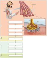
The process of muscle contraction involves several steps:
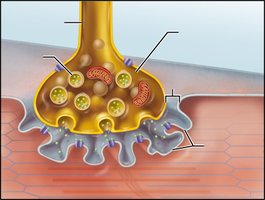
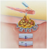
Events at the Neuromuscular Junction: Motor neuron releases acetylcholine (ACh), which binds to receptors on the sarcolemma.
Muscle Fiber Excitation: ACh binding triggers an action potential (AP) in the muscle fiber.
Excitation-Contraction Coupling: AP travels along the sarcolemma and T tubules, causing Ca2+ release from the SR.
Cross Bridge Cycling: Ca2+ binds to troponin, exposing myosin-binding sites on actin, allowing contraction.
Events at the Neuromuscular Junction
When an action potential arrives at the axon terminal:
Voltage-gated Ca2+ channels open, allowing Ca2+ entry.
Ca2+ triggers ACh release into the synaptic cleft.
ACh binds to receptors, opening Na+ channels and generating an end plate potential.
ACh is degraded by acetylcholinesterase, terminating its effect.
Generation of an Action Potential Across the Sarcolemma
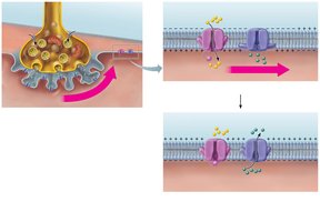
The action potential is generated and propagated in three steps:
Generation of End Plate Potential: Local depolarization due to Na+ influx.
Depolarization: If threshold is reached, voltage-gated Na+ channels open, propagating the AP.
Repolarization: Voltage-gated K+ channels open, restoring resting membrane potential.
Excitation-Contraction Coupling
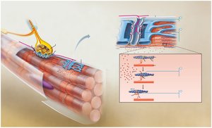
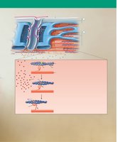
Excitation-contraction coupling links the AP to muscle contraction:
AP travels along sarcolemma and T tubules.
Voltage-sensitive proteins trigger Ca2+ release from SR.
Ca2+ binds to troponin, shifting tropomyosin and exposing myosin-binding sites.
Contraction occurs as myosin heads bind actin and cycle through cross bridge formation.
Cross Bridge Cycling
Steps of the Cross Bridge Cycle
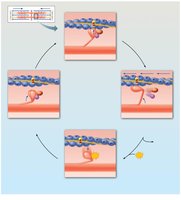
Muscle contraction is driven by the cross bridge cycle, which consists of four steps:
Cross Bridge Formation: Myosin head binds to actin.
Power Stroke: Myosin head pivots, pulling actin toward the M line.
Cross Bridge Detachment: ATP binds to myosin, causing detachment from actin.
Cocking of Myosin Head: ATP hydrolysis re-energizes the myosin head for the next cycle.
Clinical Application: Rigor Mortis
Rigor mortis is the postmortem stiffening of muscles due to the absence of ATP, preventing Ca2+ reuptake and cross bridge detachment. Muscles remain contracted until proteins degrade.
Key Terms and Concepts
ATP: Adenosine triphosphate, the energy currency for muscle contraction.
Action Potential (AP): Electrical signal that triggers muscle contraction.
Acetylcholine (ACh): Neurotransmitter at the neuromuscular junction.
Troponin and Tropomyosin: Regulatory proteins controlling access to myosin-binding sites on actin.
Na+-K+ Pump: Restores ionic conditions after AP.
Summary Table: Comparison of Muscle Types
Characteristic | Skeletal | Cardiac | Smooth |
|---|---|---|---|
Location | Attached to bones/skin | Heart | Walls of hollow organs |
Control | Voluntary | Involuntary | Involuntary |
Striations | Present | Present | Absent |
Cell Shape | Long, cylindrical | Branched | Spindle-shaped |
Nuclei | Multinucleate | 1-2 nuclei | Single nucleus |
Key Equations
Resting Membrane Potential:
Na+-K+ Pump:
ATP Hydrolysis:
Additional info: Academic context and expanded explanations were added to ensure completeness and clarity for exam preparation.
