 Indietro
Muscles and Muscle Tissue: Structure, Function, and Contraction
Guida di studio - Note intelligenti
 Appunti personalizzati basati sui tuoi materiali, ampliati con definizioni chiave, esempi e contesto.
Types of Muscle Tissue
Terminology and Classification
Muscle tissue is classified into three main types: skeletal, cardiac, and smooth. Prefixes such as myo-, mys-, and sarco- are commonly used in muscle terminology. For example, sarcoplasm refers to the cytoplasm of a muscle cell.
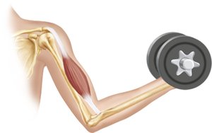
Skeletal Muscle: Attached to bones and skin, responsible for voluntary movements, characterized by long, striated fibers.
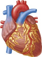
Cardiac Muscle: Found only in the heart, striated, involuntary, contracts at a steady rate due to the heart's pacemaker.
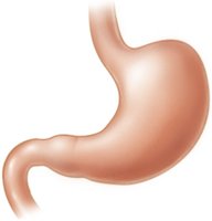
Smooth Muscle: Located in walls of hollow organs (e.g., stomach, bladder, airways), non-striated, involuntary.
Muscle Type | Location | Striations | Control |
|---|---|---|---|
Skeletal | Bones, skin | Striated | Voluntary |
Cardiac | Heart | Striated | Involuntary |
Smooth | Hollow organs | Non-striated | Involuntary |
Muscle Fiber Structure
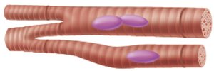
Muscle fibers are elongated cells found in skeletal and smooth muscle. Cardiac muscle cells are shorter and branched.
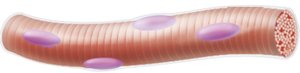
Skeletal muscle fibers: Long, cylindrical, multinucleated.
Cardiac muscle fibers: Short, branched, usually one or two nuclei.
Smooth muscle fibers: Spindle-shaped, single nucleus.

Characteristics and Functions of Muscle Tissue
Properties of Muscle Tissue
All muscle tissues share four main characteristics:
Excitability: Ability to receive and respond to stimuli.
Contractility: Ability to shorten forcibly when stimulated.
Extensibility: Ability to be stretched.
Elasticity: Ability to recoil to resting length.
Functions of Muscle Tissue
Produce movement: Responsible for locomotion and manipulation (e.g., walking, digesting, pumping blood).
Maintain posture and body position
Stabilize joints
Generate heat: As muscles contract, they produce heat.
Skeletal Muscle: Connective Tissue Sheaths
Organization of Connective Tissue Sheaths
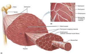
Each skeletal muscle and muscle fiber is covered by connective tissue sheaths that support and reinforce the muscle:
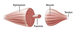
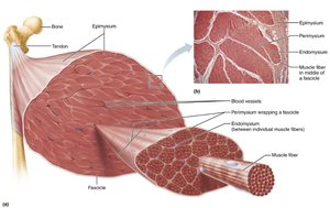
Epimysium: Dense irregular connective tissue surrounding the entire muscle.
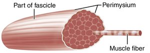
Perimysium: Fibrous connective tissue surrounding fascicles (groups of muscle fibers).
Endomysium: Fine areolar connective tissue surrounding each muscle fiber.
Structure and Organizational Levels of Skeletal Muscle
Hierarchical Organization
Skeletal muscle is organized into several structural levels:
Muscle: Entire organ, covered by epimysium.
Fascicle: Bundle of muscle fibers, covered by perimysium.
Muscle fiber: Single muscle cell, covered by endomysium.
Myofibril: Rod-like unit within muscle fiber, responsible for contraction.
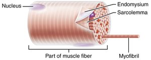
Skeletal Muscle Fiber: Microscopic Anatomy
Cellular Structure
Skeletal muscle fibers are long, cylindrical cells with multiple nuclei. The sarcolemma is the plasma membrane, and the sarcoplasm is the cytoplasm, containing glycogen and myoglobin for energy and oxygen storage.
Myofibrils: Densely packed, rodlike elements, account for ~80% of muscle cell volume.
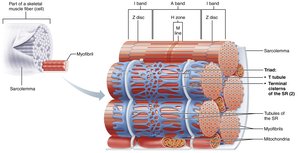
Sarcoplasmic reticulum: Specialized smooth endoplasmic reticulum for Ca2+ storage.
T tubules: Invaginations of the sarcolemma that help transmit action potentials.
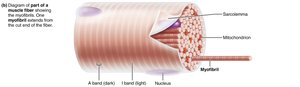
Myofibrils: Structure and Function
Striations and Sarcomeres
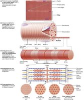
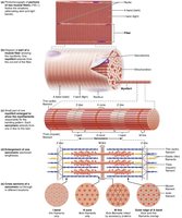
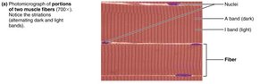
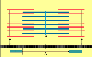
Myofibrils display striations due to the arrangement of actin (thin) and myosin (thick) filaments. The basic contractile unit is the sarcomere, defined as the region between two Z discs.
A bands: Dark regions, contain thick filaments.
I bands: Light regions, contain thin filaments.
H zone: Lighter region in the middle of the A band.
M line: Protein line bisecting the H zone.
Z disc: Coin-shaped sheet of proteins in the I band.
Myofilaments: Composition and Arrangement
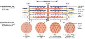
Myofilaments are organized within the sarcomere:
Actin (thin) filaments: Extend across I band and partway into A band, anchored to Z discs.
Myosin (thick) filaments: Extend length of A band, connected at M line.
Regulatory proteins: Tropomyosin and troponin complex regulate contraction.
Sarcoplasmic Reticulum and T Tubules
Calcium Regulation and Muscle Contraction
The sarcoplasmic reticulum (SR) is a network of tubules surrounding each myofibril, storing and releasing Ca2+. Terminal cisterns form cross channels at the A–I band junction. T tubules transmit action potentials deep into the muscle fiber, triggering Ca2+ release and muscle contraction.
Sliding Filament Model of Contraction
Mechanism of Muscle Contraction
According to the sliding filament theory, cross bridge attachments form and break, pulling thin filaments toward the center of the sarcomere in a ratcheting action (power stroke). This causes shortening of the muscle fiber:
I bands shorten
Z discs become closer
H zones disappear
A bands move closer together
Skeletal Muscle Contraction: Ion Channels and Neuromuscular Junction
Ion Channels
Ion channels play a major role in changing membrane potentials:
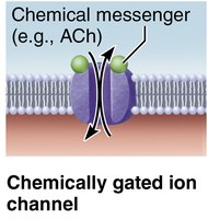
Chemically gated ion channels: Opened by neurotransmitters (e.g., acetylcholine).
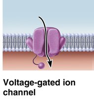
Voltage-gated ion channels: Open or close in response to voltage changes.
Neuromuscular Junction (NMJ)
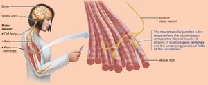
Skeletal muscles are stimulated by somatic motor neurons. The axon terminal and muscle fiber are separated by the synaptic cleft. Synaptic vesicles contain acetylcholine (ACh), which binds to receptors on the sarcolemma, triggering muscle contraction.
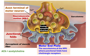
Events at the Neuromuscular Junction
Sequence of Events
Action potential arrives at axon terminal.
Voltage-gated Ca2+ channels open; Ca2+ enters motor neuron.
Ca2+ entry causes release of ACh by exocytosis.
ACh diffuses across synaptic cleft and binds to receptors on sarcolemma.
ACh binding opens ion channels, allowing Na+ in and K+ out.
Net influx of Na+ causes end plate potential (EPP).
Acetylcholinesterase degrades ACh, closing ion channels and ending contraction.
Generation of an Action Potential Across the Sarcolemma
Depolarization and Repolarization
Depolarization occurs when Na+ enters the cell, making the interior less negative. If threshold is reached, voltage-gated Na+ channels open, triggering an action potential. Repolarization restores resting conditions as K+ exits the cell and the Na+-K+ pump restores ionic balance.
Muscle Fiber Contraction: Cross Bridge Cycle
Steps of the Cross Bridge Cycle
Cross bridge formation: High-energy myosin head attaches to actin.
Power stroke: Myosin head pivots, pulling actin toward M line.
Cross bridge detachment: ATP binds to myosin, causing detachment.
Cocking of myosin head: ATP hydrolysis re-cocks myosin head.
Excitation-Contraction (E-C) Coupling
Linking Electrical and Mechanical Events
Excitation-contraction coupling describes how the action potential propagates along the sarcolemma and down T tubules, triggering Ca2+ release from the SR and initiating contraction. When stimulation stops, Ca2+ is pumped back into the SR, and the muscle relaxes.
Clinical Applications
Botulinum Toxin (Botox)
Botox is used to treat migraines, muscle spasms, and wrinkles by inhibiting ACh release, preventing muscle contraction.
Black Widow Spider Venom
Enhances ACh secretion, causing excessive muscle contraction and abdominal cramps.
Rigor Mortis
After death, ATP production ceases, Ca2+ accumulates, and cross bridges cannot detach, resulting in muscle stiffness until proteins break down.
Summary Table: Muscle Tissue Comparison
Feature | Skeletal | Cardiac | Smooth |
|---|---|---|---|
Location | Bones | Heart | Hollow organs |
Striations | Yes | Yes | No |
Control | Voluntary | Involuntary | Involuntary |
Cell shape | Long, cylindrical | Short, branched | Spindle-shaped |
Nuclei | Multinucleated | 1-2 nuclei | Single nucleus |
