 Indietro
Muscles and Muscle Tissue: Structure, Function, and Microanatomy
Guida di studio - Note intelligenti
 Appunti personalizzati basati sui tuoi materiali, ampliati con definizioni chiave, esempi e contesto.
Overview of Muscle Tissue
Introduction to Muscle Tissue
Muscle tissue comprises nearly half of the body's mass and is essential for transforming chemical energy (ATP) into mechanical energy, enabling force generation and movement. The study of muscle tissue involves understanding its types, characteristics, and functions.
Key Point 1: Muscle tissue is specialized for contraction and movement.
Key Point 2: Prefixes such as myo, mys, and sarco are commonly used in muscle terminology.
Example: Sarcoplasm refers to the cytoplasm of a muscle cell.
Types of Muscle Tissue
Skeletal Muscle
Skeletal muscle tissue is organized into muscles attached to bones and skin. These fibers are the longest, striated, and under voluntary control. Skeletal muscles contract rapidly, tire easily, and are powerful.
Key Point 1: Skeletal muscle is voluntary, striated, and attached to bones.
Key Point 2: Muscle fibers are elongated cells referred to as muscle fibers.
Example: Muscles used in athletic movements, such as throwing a discus.
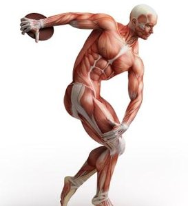
Cardiac Muscle
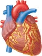
Cardiac muscle tissue is found exclusively in the heart. It is striated but involuntary, meaning it contracts without conscious control.
Key Point 1: Cardiac muscle is involuntary and striated.
Key Point 2: Cardiac muscle cells are branching and typically have one or two nuclei.
Example: The heart's rhythmic contractions pump blood throughout the body.
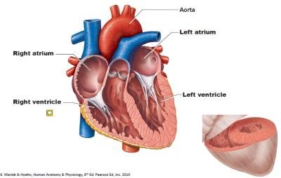
Smooth Muscle
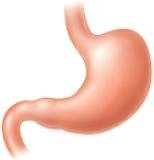
Smooth muscle tissue is located in the walls of hollow organs such as the stomach, bladder, and blood vessels. It is not striated and is involuntary.
Key Point 1: Smooth muscle is involuntary and non-striated.
Key Point 2: Cells are spindle-shaped and have a single nucleus.
Example: The detrusor muscle in the bladder wall helps expel urine.
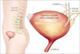
Comparison of Muscle Tissue Types
Structural and Functional Differences
The three muscle tissue types differ in location, cell shape, and appearance.
Characteristic | Skeletal | Cardiac | Smooth |
|---|---|---|---|
Body Location | Attached to bones or skin | Walls of the heart | Walls of hollow organs |
Cell Shape & Appearance | Long, cylindrical, multinucleate, striated | Branching chains, uni/binucleate, striated | Fusiform, uninucleate, no striations |
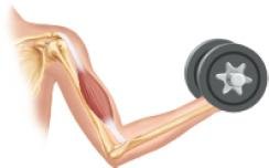
Characteristics of Muscle Tissue
Four Main Properties
All muscle tissues share four fundamental characteristics:
Excitability (Responsiveness): Ability to receive and respond to stimuli.
Contractility: Ability to shorten forcibly when stimulated.
Extensibility: Ability to be stretched.
Elasticity: Ability to recoil to resting length after stretching.
Functions of Muscle Tissue
Primary and Additional Functions
Muscles perform several vital functions:
Production of Movement: Muscles are responsible for all movements of the body.
Maintain Posture and Body Position: Muscles stabilize the body and maintain posture.
Stabilize Joints: Muscles reinforce and stabilize joints.
Generate Heat: Muscle contractions produce heat, helping maintain body temperature.
Additional Functions: Protect organs, form valves, control pupil size, and cause "goosebumps".
Skeletal Muscle Anatomy
Organ Structure and Features
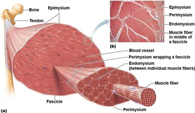
Skeletal muscle is an organ composed of multiple tissues, including nerve and blood supply, connective tissue sheaths, and attachments.
Nerve and Blood Supply: Each muscle receives a nerve, artery, and veins. Every muscle fiber is supplied by nerves, and contracting fibers require substantial oxygen and nutrients.
Connective Tissue Sheaths: Muscles and fibers are covered by connective tissue layers:
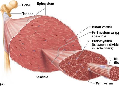
Epimysium: Surrounds the entire muscle.
Perimysium: Surrounds fascicles (bundles of muscle fibers).
Endomysium: Surrounds individual muscle fibers.
Muscle Attachments
Muscles span joints and attach to bones at two points:
Insertion: Attachment to movable bone.
Origin: Attachment to immovable or less movable bone.
Direct Attachment: Epimysium fused to periosteum or perichondrium.
Indirect Attachment: Connective tissue extends as tendon or aponeurosis.
Muscle Fiber Microanatomy
Structure of Skeletal Muscle Fibers
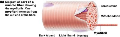
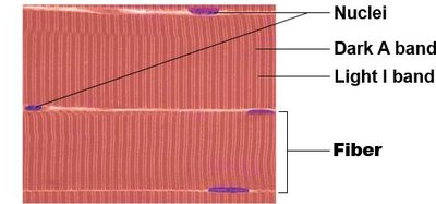
Skeletal muscle fibers are long, cylindrical cells with multiple nuclei.
Sarcolemma: Plasma membrane of the muscle fiber.
Sarcoplasm: Cytoplasm containing glycosomes and myoglobin.
Modified Organelles: Includes myofibrils, sarcoplasmic reticulum, and T tubules.
Myofibrils
Myofibrils are densely packed, rodlike elements within muscle fibers.
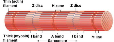
Striations: Alternating dark (A bands) and light (I bands) regions.
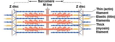
Sarcomeres: Smallest contractile units, aligned end-to-end.
Myofilaments: Actin (thin) and myosin (thick) filaments arranged within sarcomeres.
Molecular Composition of Myofilaments
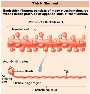
Thick Filaments: Composed of myosin protein. Myosin heads form cross bridges during contraction.
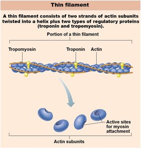
Thin Filaments: Composed of actin, with regulatory proteins tropomyosin and troponin. Tropomyosin blocks myosin binding sites at rest; troponin binds calcium and moves tropomyosin to allow contraction.
Sarcoplasmic Reticulum and T Tubules
Calcium Storage and Electrical Transmission
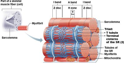
Sarcoplasmic Reticulum (SR): Network of smooth endoplasmic reticulum surrounding each myofibril, storing and releasing calcium.
T Tubules: Extensions of the sarcolemma that transmit electrical impulses deep into the muscle fiber.
Triad: Structure formed by a T tubule and two terminal cisterns of the SR.
Case Study: Muscle Injury and Healing
Muscle Stability and Injury
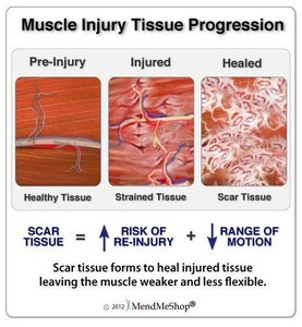
Muscles contribute to joint stability. Injuries such as hamstring strains affect the joints they span and involve tissue healing.
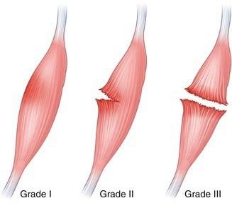
Key Point 1: Muscle injuries can be classified by severity (Grade I, II, III).
Key Point 2: Healing involves development of scar tissue, which may reduce flexibility and increase risk of re-injury.
Optimal Healing: Requires proper tissue regeneration, rehabilitation, and prevention of excessive scar formation.
Summary Table: Muscle Tissue Types
Type | Location | Control | Striations | Cell Shape |
|---|---|---|---|---|
Skeletal | Bones, skin | Voluntary | Yes | Long, cylindrical, multinucleate |
Cardiac | Heart | Involuntary | Yes | Branching, uni/binucleate |
Smooth | Hollow organs | Involuntary | No | Fusiform, uninucleate |
Key Equations and Concepts
ATP and Muscle Contraction
Muscle contraction is powered by ATP hydrolysis:
Equation:
Application: Energy released is used for cross-bridge cycling and muscle contraction.
Summary
Understanding muscle tissue types, their structure, and function is fundamental to anatomy and physiology. Skeletal, cardiac, and smooth muscles each play unique roles in movement, stability, and organ function. Injury and healing processes are important for clinical and athletic contexts. Additional info: Academic context was added to clarify muscle injury grades and healing processes.
